- Ciliary body
-
Ciliary body 
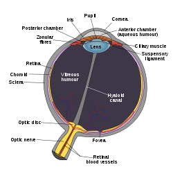
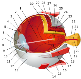
Schematic diagram of the human eye Latin corpus ciliare Gray's subject #225 1010 Artery long posterior ciliary arteries MeSH Ciliary+Body The ciliary body is the circumferential tissue inside the eye composed of the ciliary muscle and ciliary processes.[1] It is triangular in horizontal section and is coated by a double layer, the ciliary epithelium. This epithelium produces the aqueous humor.[2] The inner layer is transparent and covers the vitreous body, and is continuous from the neural tissue of the retina. The outer layer is highly pigmented, continuous with the retinal pigment epithelium, and constitutes the cells of the dilator muscle. This double membrane is often regarded to be continuous with the retina and a rudiment of the embryological correspondent to the retina. The inner layer is unpigmented until it reaches the iris, where it takes on pigment. The retina ends at the ora serrata. It is part of the uveal tract— the layer of tissue which provides most of the nutrients in the eye. It extends from the ora serrata to the root of the iris. There are three sets of ciliary muscles in the eye, the longitudinal, radial, and circular muscles. They are near the front of the eye, above and below the lens. They are attached to the lens by connective tissue called the zonule of Zinn, and are responsible for shaping the lens to focus light on the retina. The ciliary body receives parasympathetic innervation from the oculomotor nerve.
Contents
Functions
The ciliary body has three functions: accommodation, aqueous humor production and the production and maintenance of the lens zonules. It also anchors the lens in place. Accomommodation essentially means that when the ciliary muscle contracts, the lens becomes more convex, generally improving the focus for closer objects. When it relaxes it flattens the lens, generally improving the focus for farther objects. One of the essential roles of the ciliary body is also the production of the aqueous humor, which is responsible for providing most of the nutrients for the lens and the cornea and involved in waste management of these areas.
Clinical significance
The ciliary body is the main target of drugs against glaucoma (apraclonidine) as it is responsible for aqueous humor production. Its inhibition leads to the lowering of aqueous humor production and causes a subsequent drop in the intraocular pressure.
See also
References
External links
- Ciliary+body at eMedicine Dictionary
- Histology at BU 08011loa
- Atlas of anatomy at UMich eye_1 - "Sagittal Section Through the Eyeball"
Sensory system – visual system – globe of eye (TA A15.2.1–6, TH 3.11.08.0-5, GA 10.1005) Fibrous tunic (outer) Uvea/vascular tunic (middle) Ciliary bodyRetina (inner) LayersCellsPhotoreceptor cells (Cone cell, Rod cell) → (Horizontal cell) → Bipolar cell → (Amacrine cell) → Retina ganglion cell (Midget cell, Parasol cell, Bistratified cell, Giant retina ganglion cells, Photosensitive ganglion cell) → Diencephalon: P cell, M cell, K cell
Muller gliaOtherAnterior segment Posterior segment Other M: EYE
anat(g/a/p)/phys/devp/prot
noco/cong/tumr, epon
proc, drug(S1A/1E/1F/1L)
Categories:- Eye anatomy
Wikimedia Foundation. 2010.


