- Oculomotor nerve
-
Nerve: Oculomotor nerve 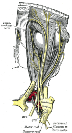
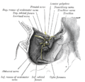
Nerves of the orbit. Seen from above. 
Inferior view of the human brain, with the cranial nerves labelled. Latin nervus oculomotorius Gray's subject #198 884 Innervates Superior rectus, Inferior rectus, Medial rectus, Inferior oblique, Levator palpebrae, sphincter pupillae (parasympathetics), ciliaris muscle (parasympathetics) From oculomotor nucleus, Edinger-Westphal nucleus To superior branch, inferior branch MeSH Oculomotor+Nerve Cranial Nerves CN I – Olfactory CN II – Optic CN III – Oculomotor CN IV – Trochlear CN V – Trigeminal CN VI – Abducens CN VII – Facial CN VIII – Vestibulocochlear CN IX – Glossopharyngeal CN X – Vagus CN XI – Spinal Accessory CN XII – Hypoglossal The oculomotor nerve is the 3rd of 12 paired cranial nerves. It controls most of the eye's movement and constriction of the pupil, and maintains an open eyelid. The optic nerve is derived from the basal plate of the embryonic midbrain. Cranial nerves IV and VI also participate in control of eye movement.
Contents
Path
Nuclei
The oculomotor nerve (CN III) arises from the anterior aspect of mesencephalon (midbrain). There are two nuclei for the oculomotor nerve:
- The oculomotor nucleus originates at the level of the superior colliculus. The muscles it controls are the striated muscle in levator palpebrae superioris and all extraocular muscles except for the superior oblique muscle and the lateral rectus muscle.
- The Edinger-Westphal nucleus supplies parasympathetic fibres to the eye via the ciliary ganglion, and thus controls the sphincter pupillae muscle (affecting pupil constriction) and the ciliary muscle (affecting accommodation).
Sympathetic postganglionic fibres also join the nerve from the plexus on the internal carotid artery in the wall of the cavernous sinus and are distributed through the nerve, e.g., to the smooth muscle of levator palpebrae superioris.
Emergence from brain
On emerging from the brain, the nerve is invested with a sheath of pia mater, and enclosed in a prolongation from the arachnoid.
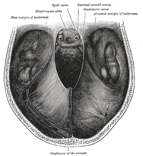
It passes between the superior cerebellar (below) and posterior cerebral arteries (above), and then pierces the dura mater anterior and lateral to the posterior clinoid process, passing between the free and attached borders of the tentorium cerebelli.
It runs along the lateral wall of the cavernous sinus, above the other orbital nerves, receiving in its course one or two filaments from the cavernous plexus of the sympathetic, and a communicating branch from the ophthalmic division of the trigeminal.
Superior and inferior rami
It then divides into two branches, which enter the orbit through the superior orbital fissure, between the two heads of the lateral rectus.
Here the nerve is placed below the trochlear nerve and the frontal and lacrimal branches of the ophthalmic nerve, while the nasociliary nerve is placed between its two rami:
Testing the oculomotor nerve
Eye muscles
Cranial nerves III, IV, and VI are usually tested together. The examiner typically instructs the patient to hold his head still and follow only with the eyes a finger or penlight that circumscribes a large "H" in front of the patient. By observing the eye movement and eyelids, the examiner is able to obtain more information about the extraocular muscles, the levator palpebrae superioris muscle, and cranial nerves III, IV, and VI.
Since the oculomotor nerve controls most of the eye muscles, it may be easier to detect damage to it. Damage to this nerve, termed oculomotor nerve palsy is also known by the down n' out symptoms, because of the position of the affected eye.
Pupillary reflex
The oculomotor nerve also controls the constriction of the pupils and thickening of the lens of the eye. This can be tested in two main ways. By moving a finger toward a person's face to induce accommodation, as well as his going cross-eyed, his pupils should constrict.
Shining a light into one eye should result in equal constriction of the other eye. The neurons in the optic nerve decussate in the optic chiasm with some crossing to the contralateral optic nerve tract. This is the basis of the "swinging-flashlight test".
Pathology
Paralysis of the oculomotor nerve, i.e., oculomotor nerve palsy, is a rare condition. It can arise due to:
- direct trauma,
- demyelinating diseases (e.g., multiple sclerosis),
- increased intracranial pressure (leading to uncal herniation)
- due to a space-occupying lesion (e.g., brain cancer) or a
- spontaneous subarachnoid haemorrhage (e.g., berry aneurysm), and
- microvascular disease, e.g., diabetes.
In people with diabetes and older than 50 years of age, an oculomotor nerve palsy, in the classical sense, occurs with sparing (or preservation) of the pupillary reflex. This is thought to arise due the anatomical arrangement of the nerve fibers in the oculomotor nerve; fibers controlling the pupillary function are superficial and spared from ischemic injuries typical of diabetes. On the converse, a subarachnoid haemorrhage, which leads to compression of the oculomotor nerve, usually affects the superficial fibers and manifests as a palsy with loss of the pupillary reflex.[1]
Additional images
-
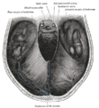
Dura mater and its processes exposed by removing part of the right half of the skull, and the brain.
-
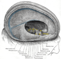
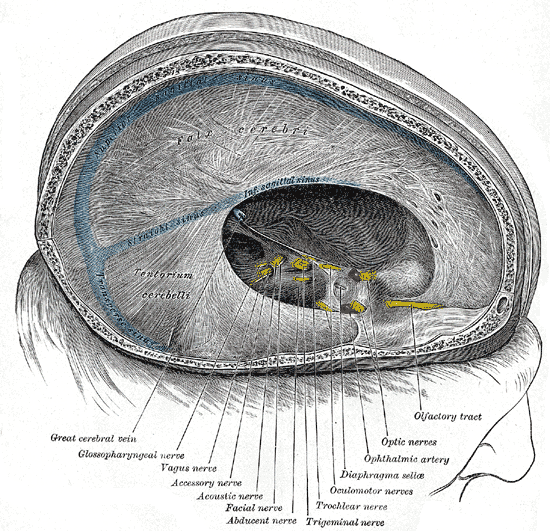
Tentorium cerebelli from above.
-
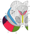
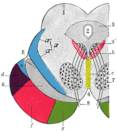
Coronal section through mid-brain.
-
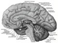
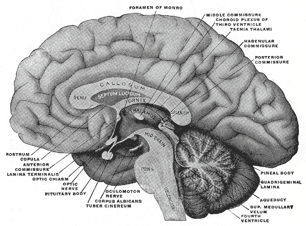
Mesal aspect of a brain sectioned in the median sagittal plane.
-
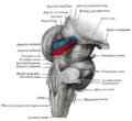
Hind- and mid-brains; postero-lateral view.
-

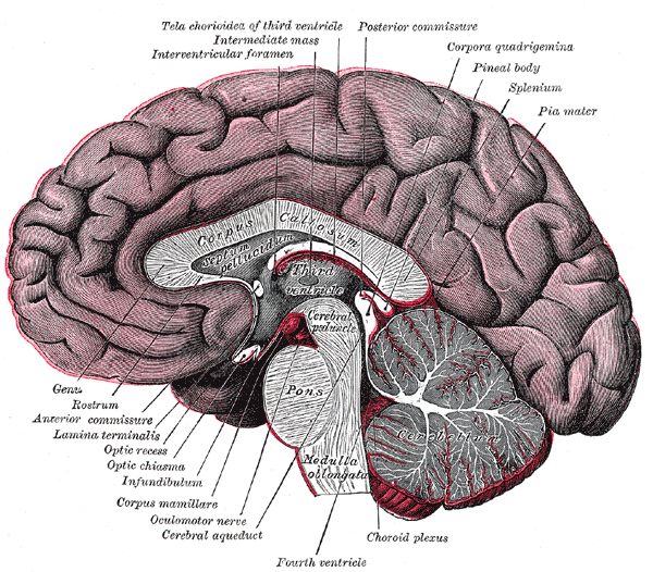
Median sagittal section of brain.
-
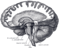
Dissection showing the course of the cerebrospinal fibers.
-

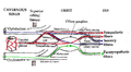
Plan of oculomotor nerve.
-

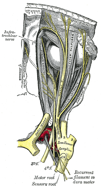
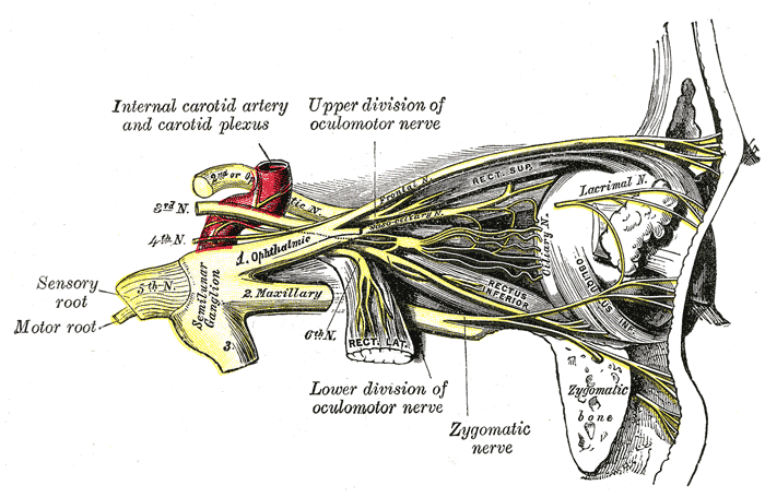
Nerves of the orbit, and the ciliary ganglion. Side view.
-

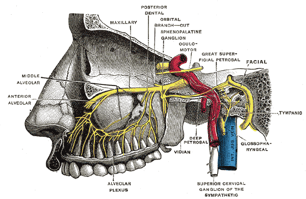
Alveolar branches of superior maxillary nerve and sphenopalatine ganglion.
-

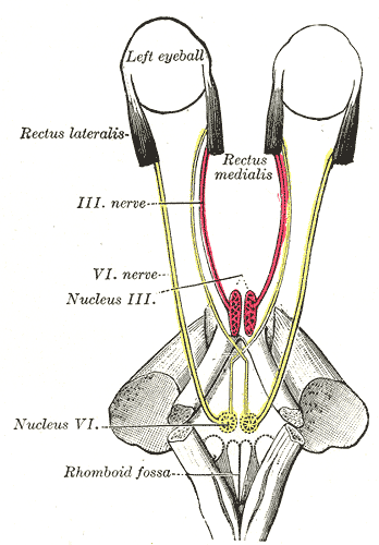
Figure showing the mode of innervation of the Recti medialis and lateralis of the eye.
-
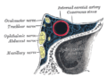
Oblique section through the right cavernous sinus.
-
Dissection showing origins of right ocular muscles, and nerves entering by the superior orbital fissure.
-
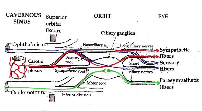
Pathways in the Ciliary Ganglion.
References
- ^ Goodwin J. Oculomotor Nerve Palsy. eMedicine.com. URL:http://emedicine.medscape.com/article/1198462-overview. Accessed on: January 16, 2009.
See also
External links
- MedEd at Loyola GrossAnatomy/h_n/cn/cn1/cn3.htm
- oph/183 at eMedicine - "Oculomotor nerve palsy"
- MeSH Oculomotor+Nerve
- NeuroNames hier-479
- cranialnerves at The Anatomy Lesson by Wesley Norman (Georgetown University) (III)
- Notes on Oculomotor Nerve
Nerves of head and neck: the cranial nerves and nuclei (TA A14.2.01, GA 9.855) olfactory (AON->I) optic (LGN->II) oculomotor
(ON, EWN->III)trochlear (TN->IV) no significant branchestrigeminal
(PSN, TSN, MN, TMN->V)abducens (AN->VI) no significant branchesfacial (FMN, SN, SSN->VII) near origininside
facial canalvestibulocochlear
(VN, CN->VIII)glossopharyngeal
(NA, ISN, SN->IX)before jugular fossaafter jugular fossavagus
(NA, DNVN, SN->X)before jugular fossaafter jugular fossaaccessory (NA, SAN->XI) hypoglossal (HN->XII) Categories:


Wikimedia Foundation. 2010.