- Mandibular nerve
-
Nerve: Mandibular nerve 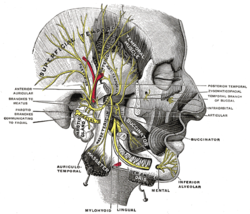
Mandibular division of the trigeminal nerve. 
Mandibular division of trigeminal nerve, seen from the middle line. The small figure is an enlarged view of the otic ganglion. Latin n. mandibularis Gray's subject #200 893 From trigeminal nerve MeSH Mandibular+Nerve The mandibular nerve (V3) is the largest of the three branches of the trigeminal nerve.
Contents
Structure
Roots
It is made up of two roots:
- a large sensory root proceeding from the inferior angle of the trigeminal ganglion.
- a small motor root (the motor part of the trigeminal), which passes beneath the ganglion, and unites with the sensory root, just after its exit through the foramen ovale.
Path
The two roots (sensory and motor) exit the middle cranial fossa through the foramen ovale. The two roots then combine.
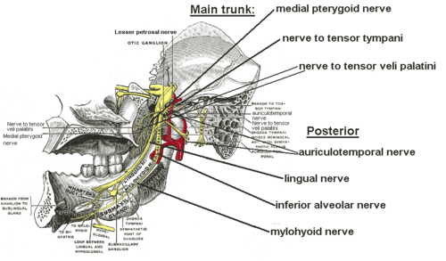
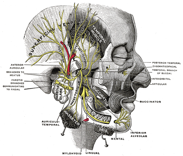
Immediately in the infratemporal fossa beneath the base of the skull, the nerve gives off two branches from its medial side: a recurrent branch (nervus spinosus) and the nerve to the medial pterygoid muscle. The mandibular nerve then divides into two trunks, an anterior and a posterior.
Branches
 Branches from the main trunk (except nervus spinosus) and the posterior division.
The mandibular nerve gives off the following branches:
- From the main trunk of the nerve (before the division)
- nervus spinosus (meningeal branch)
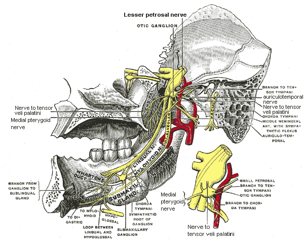
- medial pterygoid nerve
- nerve to tensor tympani
- nerve to tensor veli palatini
- From the anterior division
- masseteric nerve
- deep temporal nerves (anterior and posterior)
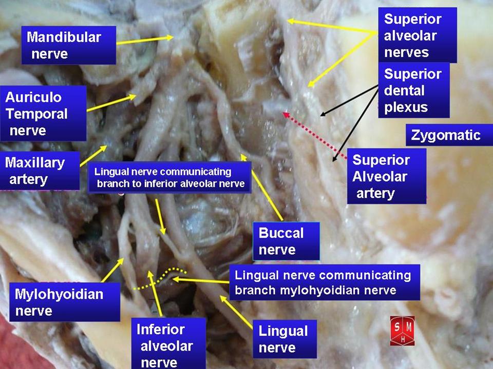
- buccal nerve (a sensory nerve)
- lateral pterygoid nerve
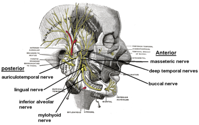
- From the posterior division
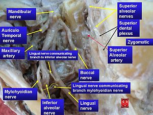
- auriculotemporal nerve
- lingual nerve
- inferior alveolar nerve
- motor branch to mylohyoid and anterior belly of digastric muscles (mylohyoid nerve)
The mandibular nerve also gives off branches to the otic ganglion
Supplies
The mandibular nerve innervates:
- mylohyoid muscle and anterior belly of digastric muscle
- mucous membrane of the anterior two-thirds of the tongue
- the inside of the cheek (the buccal mucosa)
- teeth and mucoperiosteum of mandibular teeth
- skin of the temporal region
- auricula
- lower lip, and chin
- Muscles of mastication
- the muscles tensor tympani and tensor veli palatini
 Mandibular nerve
See also
Additional images
-

Dermatome distribution of the trigeminal nerve
-
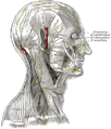
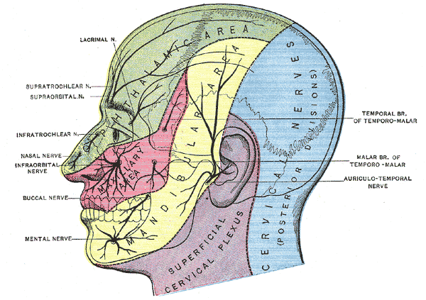
The nerves of the scalp, face, and side of neck.
-

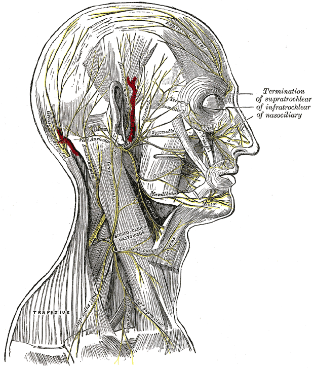
Mandibular nerve
External links
- MedEd at Loyola GrossAnatomy/h_n/cn/cn1/cnb3.htm
- SUNY Figs 27:03-02
- cranialnerves at The Anatomy Lesson by Wesley Norman (Georgetown University) (V)
Nerves of head and neck: the cranial nerves and nuclei (TA A14.2.01, GA 9.855) olfactory (AON->I) optic (LGN->II) oculomotor
(ON, EWN->III)trochlear (TN->IV) no significant branchestrigeminal
(PSN, TSN, MN, TMN->V)abducens (AN->VI) no significant branchesfacial (FMN, SN, SSN->VII) near originvestibulocochlear
(VN, CN->VIII)glossopharyngeal
(NA, ISN, SN->IX)before jugular fossaafter jugular fossavagus
(NA, DNVN, SN->X)before jugular fossaafter jugular fossaaccessory (NA, SAN->XI) hypoglossal (HN->XII) The cranial nerves: trigeminal nerve ophthalmic
(V1)frontal: supratrochlear · supraorbital (lateral branch, medial branch)
nasociliary: long ciliary · infratrochlear · posterior ethmoidal · anterior ethmoidal (external nasal, internal nasal) · sensory root of ciliary ganglion (ciliary ganglion)
lacrimalmaxillary
(V2)in meningeszygomatic (zygomaticotemporal, zygomaticofacial) · pterygopalatine (pterygopalatine ganglion see below for details) · posterior superior alveolaron facemandibular
(V3)in meningesanteriorposteriorCategories:


Wikimedia Foundation. 2010.