- Mandibular foramen
-
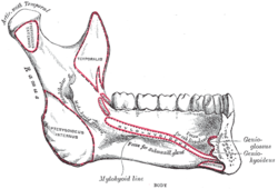
Bone: Mandibular foramen Mandible. Inner surface. Side view. (Mandibular foramen visible at left.) Latin foramen mandibulae Gray's subject #44 173 The Mandibular foramen is an opening on the internal surface of the ramus (posterior and perpendicularly oriented part of the mandible) for divisions of the mandibular vessels and nerve to pass.
Contents
Contents
The mandibular nerve is one of three branches of the trigeminal nerve (CN V) and the only branch with motor innervation.
One branch of it, the inferior alveolar nerve as well as the inferior alveolar artery enter the foramen traveling through the body and exit at the mental foramen on the anterior mandible at which point the nerve is known as the mental nerve.
These nerves provide sensory innervation to the lower teeth, as well as the lower lip and some skin on the lower face.
Structures of rim
There are two distinct anatomies to its rim.
- In the common form the rim is “V” shaped, with a groove separating the anterior and posterior parts.
- In the horizontal-oval form there is no groove, and the rim is horizontally oriented and oval in shape, the anterior and posterior parts connected.
Additional images
External links
- SUNY Labs 34:st-0211
- Photo at unc.edu
- Mandibular+foramen at eMedicine Dictionary
- cranialnerves at The Anatomy Lesson by Wesley Norman (Georgetown University) (V)
Bones of head and neck: the facial skeleton of the skull (TA A02.1.08–15, GA 2.156–177) Maxilla SurfacesProcessesOtherZygomatic Palatine FossaePlatesProcessesMandible external surface (Symphysis menti, Lingual foramen, Mental protuberance, Mental foramen, Mandibular incisive canal) · internal surface (Mental spine, Mylohyoid line, Sublingual fovea, Submandibular fovea) · Alveolar part of mandibleMylohyoid groove (Mandibular canal, Lingula) · Mandibular foramen · Angle
Coronoid process · Mandibular notch · Condyloid process · Pterygoid foveaMinor/
noseNasal bone: Internasal suture · Nasal foramina
Inferior nasal concha: Ethmoidal process · Maxillary process
Vomer: Vomer anterior · Synostosis vomerina · Vomer posterior (Wing)
Lacrimal: Posterior lacrimal crest · Lacrimal groove · Lacrimal hamulusCategories:- Foramina of the skull
- Musculoskeletal system stubs
Wikimedia Foundation. 2010.


