- Foramen spinosum
Infobox Bone
Name = Foramen spinosum of Sphenoid
Latin =
GraySubject = 35
GrayPage = 150

Caption =Sphenoid bone . Upper surface. (foramen spinosum labeled left, second from bottom.)
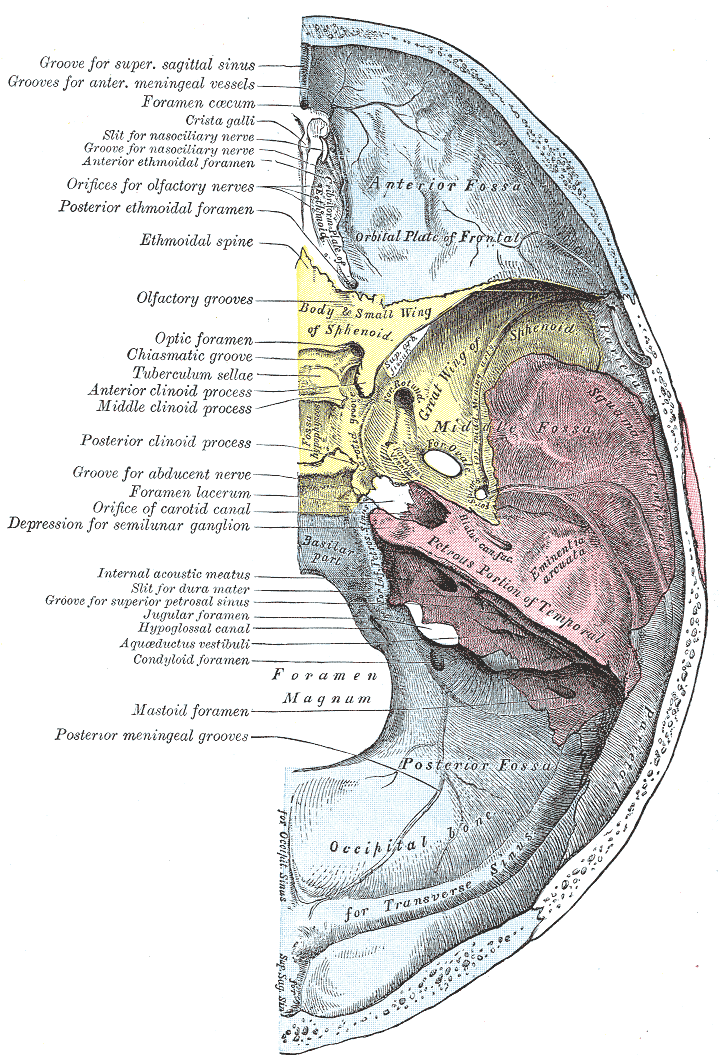
Caption2 = Base of the skull. Upper surface. (Sphenoid is yellow, and foramen spinosum is at bottom right of sphenoid.)
MeshName =
MeshNumber =
DorlandsPre = f_12
DorlandsSuf = 12373713
The foramen spinosum is one of several foramina located in the base of theskull , on thesphenoid bone , situated lateral to the foramen ovale, in a posterior angle.Contents
It permits the passage of certain arteries, veins and/or other structures:
* the
middle meningeal artery
* a recurrent branch, thenervus spinosus , from themandibular nerve (the mandibular nerve is the third branch (V3) of thetrigeminal nerve )Morphology and morphometry
The foramen may be absent (in approx. 2% of the cases), in which case the middle meningeal artery enters the
cranial cavity through the foramen ovale. cite web | title=Illustrated Encyclopedia of Human Anatomic Variation: Opus V: Skeletal Systems: Cranium - Sphenoid Bone | work=Illustrated Encyclopedia of Human Anatomic Variation | url=http://www.anatomyatlases.org/AnatomicVariants/SkeletalSystem/Text/SphenoidBone.shtml | accessdate=2006-04-10]The foramen spinosum and ovale may be continuous, and the foramen spinosum may be duplicated. Wood-Jones (1931) found the foramen spinosum to be more or less incomplete in approx. 44% of the cases. The foramen spinosum was small or altogether absent in 0.4% of Lindblom's (1936) cases. This is especially true when the middle meningeal artery arises from the
ophthalmic artery (the foramen would be near to empty in that case). In rare cases, early division of the middle meningeal artery into a posterior and anterior division may result in a duplication of the foramen spinosum. cite web | title=Illustrated Encyclopedia of Human Anatomic Variation: Opus V: Skeletal Systems: Cranium - Sphenoid Bone | work=Illustrated Encyclopedia of Human Anatomic Variation | url=http://www.anatomyatlases.org/AnatomicVariants/SkeletalSystem/Text/SphenoidBone.shtml | accessdate=2006-04-10]In the newborn, the foramen spinosum is about 2.25 mm and in the adults about 2.56 mm in length. The width of the foramen extends from 1.05 mm to about 2.1 mm in the adults. cite journal | author=Lang J, Maier R, Schafhauser O | title=Postnatal enlargement of the foramina rotundum, ovale et spinosum and their topographical changes | journal=Anatomischer Anzeiger | year=1984 | pages=351–87 | volume=156 | issue=5 | pmid=6486466] The average diameter of the foramen spinosum is 2.63 mm in the adult. cite journal | author=Yanagi S | title=Developmental studies on the foramen rotundum, foramen ovale and foramen spinosum of the human sphenoid bone | journal=The Hokkaido Journal of Medical Science | year=1987 | pages=485–96 | volume=62 | issue=3 | pmid=3610040]
The earliest perfect ring-shaped formation of the foramen spinosum was observed in the 8th month after birth and the latest in 7 years after birth in a developmental study on the
foramen rotundum , foramen ovale and foramen spinosum. The majority of the foramen in the skulls studies was round in shape. cite journal | author=Yanagi S | title=Developmental studies on the foramen rotundum, foramen ovale and foramen spinosum of the human sphenoid bone | journal=The Hokkaido Journal of Medical Science | year=1987 | pages=485–96 | volume=62 | issue=3 | pmid=3610040] Ginsberg "et al." (see reference below) observed asymmetry of size in 16% of their patients.In a study under 123 CT studies, Ginsberg, Pruett, Chen and Elster did not find an inverse relationship between the size of the foramen spinosum and that of the foramen ovale (for instance, a smaller foramen spinosum did not correlate with the size of the foramen ovale). cite journal | author=Ginsberg LE, Pruett SW, Chen MY, Elster AD | title=Skull-base foramina of the middle cranial fossa: reassessment of normal variation with high-resolution CT | journal=Americal Journal of Neuroradiology | month=February | year=1994 | pages=283–91 | volume=15 | issue=2 | pmid=8192074]
References
ee also
*
Foramina of skull
=AdditionalExternal links
* - "Osteology of the Skull: Internal Surface of Skull"
* - "Schematic view of key landmarks of the infratemporal fossa."
*
*
* [http://zemlin.shs.uiuc.edu/Skull/slide-Pages/10.htm Anatomy of the Skull - 27. Foramen spinosum]
Wikimedia Foundation. 2010.