- Foramen ovale (skull)
Infobox Bone
Name = Foramen ovale of Sphenoid
Latin = foramen ovale ossis sphenoidalis
GraySubject = 35
GrayPage = 150

Caption =Sphenoid bone . Upper surface. (foramen ovale labeled at left, third from bottom)
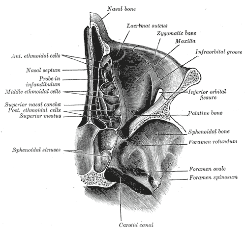
Caption2 = Horizontal section of nasal and orbital cavities.
MeshName =
MeshNumber =
DorlandsPre = f_12
DorlandsSuf = 12373448
At thebase of the skull the foramen ovale (Latin: oval window) is one of the larger of the several holes (theforamina ) that transmit nerves through the skull. The foramen ovale is situated in the anterior part of thesphenoid bone , posteriolateral to theforamen rotundum .Contents
Several nerves, arteries and veins pass through the foramen ovale. They are as follows:
*
Lesser superficial petrosal nerve of (CN IX) (note: the lesser superficial petrosal nerve sometimes passes through a special canal ("canaliculus innominatus" of Arnold), situated medial to theforamen spinosum )
*Accessory meningeal artery (small meningeal or parvidural branch, sometimes derived from themiddle meningeal artery )
*Mandibular nerve (the third branch (V3) of thetrigeminal nerve )
*Emissary veins (from thecavernous sinus to thepterygoid plexus )The contents of this foramen neatly form the mnemonic 'LAME'.
The
otic ganglion is situated directly under the foramen, but is also transmitted through the foramen ovale.Morphology and morphometry
Similar to other foramina, the foramen ovale differs in shape and size throughout the natural life. The earliest perfect ring-shaped formation of the foramen ovale was observed in the 7th fetal month and the latest in 3 years after birth, in a study using over 350 skulls. cite journal | author=Yanagi S | title=Developmental studies on the foramen rotundum, foramen ovale and foramen spinosum of the human sphenoid bone | journal=The Hokkaido Journal of Medical Science | year=1987 | pages=485–96 | volume=62 | issue=3 | pmid=3610040]
In a study conducted on 100 skulls, the foramen ovale was divided into 2 or 3 components in 4.5% of the cases. The borders of the foramen in some skulls were also irregular and rough. This may suggest, based on radiological images, the presence of morbid changes, which might be the sole anatomical variation in the foramina ovale of humans. (Reymond et al.) cite journal | author=Reymond J, Charuta A, Wysocki J | title=The morphology and morphometry of the foramina of the greater wing of the human sphenoid bone | journal=Folia Morphologica | year=2005 | pages=188–93 | volume=64 | issue=3 | pmid=16228954]In newborn, the foramen ovale is about 3.85 mm and in the adults about 7.2 mm in length. The average maximal length is about 7.48 mm and its average minimal length is 4.17 mm in the adult. The width extends from 1.81 mm in the newborn to 3.7 mm in adults.cite journal | author=Yanagi S | title=Developmental studies on the foramen rotundum, foramen ovale and foramen spinosum of the human sphenoid bone | journal=The Hokkaido Journal of Medical Science | year=1987 | pages=485–96 | volume=62 | issue=3 | pmid=3610040] cite journal | author=Lang J, Maier R, Schafhauser O | title=Postnatal enlargement of the foramina rotundum, ovale et spinosum and their topographical changes | journal=Anatomischer Anzeiger | year=1984 | pages=351–87 | volume=156 | issue=5 | pmid=6486466]
=AdditionalReferences
ee also
*
Foramina of skull External links
*
* (NormanAnatomyFig|IX)
*
*
* [http://zemlin.shs.uiuc.edu/Skull/slide-Pages/10.htm Adult skull as seen from beneath at uiuc.edu]
Wikimedia Foundation. 2010.