- Orbit (anatomy)
-
Orbit (anatomy) 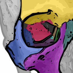
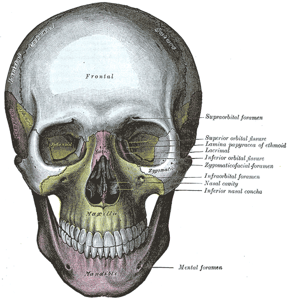
The seven bones that articulate to form the orbit.
yellow = Frontal bone
green = Lacrimal bone
brown = Ethmoid bone
blue = Zygomatic bone
purple = Maxillary bone
aqua = Palatine bone
red = Sphenoid bone
teal = Nasal bone
The nasal bone is illustrated but does not form part of the orbitLatin orbitae Gray's subject #46 188 MeSH Orbit In anatomy, the orbit is the cavity or socket of the skull in which the eye and its appendages are situated. "Orbit" can refer to the bony socket,[1] or it can also be used to imply the contents.[2] In the adult human, the volume of the orbit is 30 ml, of which the eye occupies 6.5 ml.[3]
Contents
Definition
The orbits are conical or four-sided pyramidal cavities, which open into the midline of the face and point back into the head. Each consists of a base, an apex and four walls. They protect the eye from mechanical injury.[4]
The base, which opens in the face, has four borders. The following bones take part in their formation:
- Superior margin: frontal bone
- Inferior margin: maxilla and zygomatic
- Medial margin: frontal, lacrimal and maxilla
- Lateral margin: zygomatic and frontal
The apex lies near the medial end of superior orbital fissure and contains the optic canal which communicates with middle cranial fossa.
The roof (superior wall) is formed by the orbital plate frontal bone and the lesser wing of sphenoid. The orbital surface presents medially by trochlear fovea and laterally by lacrimal fossa
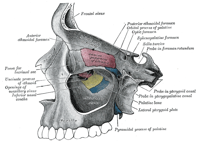
The floor (inferior wall) is formed by the orbital surface of maxilla, the orbital surface of zygomatic bone and the orbital process of palatine bone. Medially near the orbital margin is located the groove for nasolacrimal duct. Near the middle of the floor, located infraorbital groove, which leads to the infraorbital foramen. The floor is separated from the lateral wall by inferior orbital fissure, which connects the orbit to pterygopalatine and infratemporal fossa.
The medial wall is formed by the frontal process of maxilla, lacrimal bone, orbital plate of ethmoid and a small part of the body of the sphenoid.
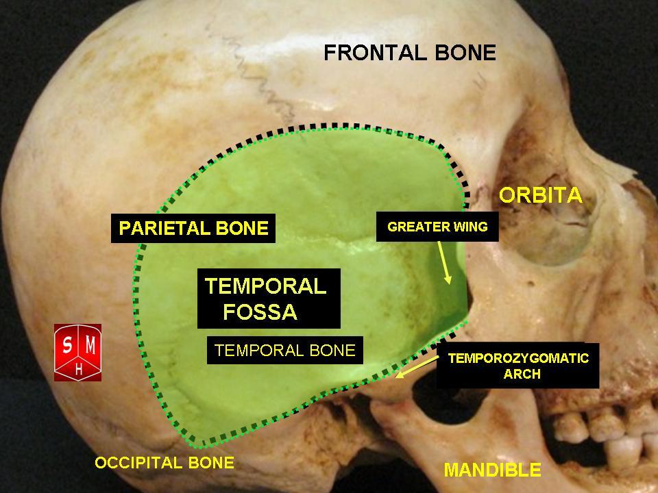
The Lateral wall is formed by the orbital process of zygomatic and the orbital plate of greater wing of sphenoid. The bones meet at the zygomaticosphenoid suture. The lateral wall is the thickest wall of the orbit. The optic foramen, which contains the optic nerve and the large ophthalmic artery, is at the nasal side of the apex, while a larger entry, the superior orbital fissure, through which veins, motor nerves, and non-visual sensory nerves (e.g., those for pain), among other fissures.
Protrusion
In the orbit, fat tissue, which surrounds the eyeball and its muscles, keeps the rotation smooth with respect to the center of the eye. If excess liquid is collected in the fat cushion tissue, the eye may protrude (known also as exophthalmos). Alternately, the eye may make an illusion of protrusion in extreme fear, not from the contraction of smooth muscle of the orbit, but based on the widening of the eyelids and dilation of the pupil (all commanded by the sympathetic nervous system.[5]
 Tear system:
a. tear gland / lacrimal gland,
b. superior lacrimal punctum,
c. superior lacrimal canal,
d. tear sac / lacrimal sac,
e. inferior lacrimal punctum,
f. inferior lacrimal canal,
g. nasolacrimal canalEnlargement of the lacrimal gland, located superotemporally within the orbit, produces protrusion of the eye inferiorly and medially (away from the location of the lacrimal gland). Lacrimal gland may be enlarged from inflammation (e.g. sarcoid) or neoplasm (e.g. lymphoma or adenoid cystic carcinoma).[6]
Tumors (e.g. glioma and meningioma of the optic nerve) within the cone formed by the horizontal rectus muscles produce axial protrusion (bulging forward) of the eye.
Graves disease may also cause axial protrusion of the eye, known as Graves' ophthalmopathy, due to buildup of extracellular matrix proteins and fibrosis in the rectus muscles. Development of Graves' ophthalmopathy may be independent of thyroid function.[7]
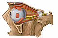
Contents
- Eyeball
- Fascias: Orbital, Bulbar
- Extraocular muscles (Levator Palpebrae Superioris, Superior, Inferior, Lateral and Medial Rectus muscles, Superior and Inferior Oblique Muscles)
- Nerves: cranial nerves II, III, IV, V, and VI
- Blood vessels
- Extraocular Fat
- Lacrimal gland, Lacrimal sac, Nasolacrimal duct
- Eyelids
- Medial palpebral ligament and Lateral palpebral ligament
- Medial and Lateral Check ligaments
- Suspensory ligament of the eyeball
- Conjunctiva
- Trochlea of superior oblique
- Orbital septum
- Ciliary ganglion and short ciliary nerves
Bones
In humans, seven bones make up the bony orbit:
- Frontal bone (Pars orbitalis)
- Lacrimal bone
- Ethmoid bone (Lamina papyracea)
- Zygomatic bone (Orbital process of the zygomatic bone)
- Maxillary bone (Orbital surface of the body of the maxilla)
- Palatine bone (Orbital process of palatine bone)
- Sphenoid bone (Greater and lesser wings)
Foramina and openings
- Optic canal
- Superior orbital fissure
- Inferior orbital fissure
- Anterior ethmoidal foramen
- Posterior ethmoidal foramen
- Infraorbital foramen
- Supraorbital foramen
- Naso-lacrimal canal opening
- Zygomatic orbital foramen
Additional images
-
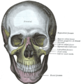
The skull from the front
-
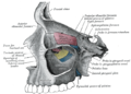
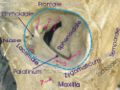
Medial wall of left orbit
-
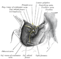
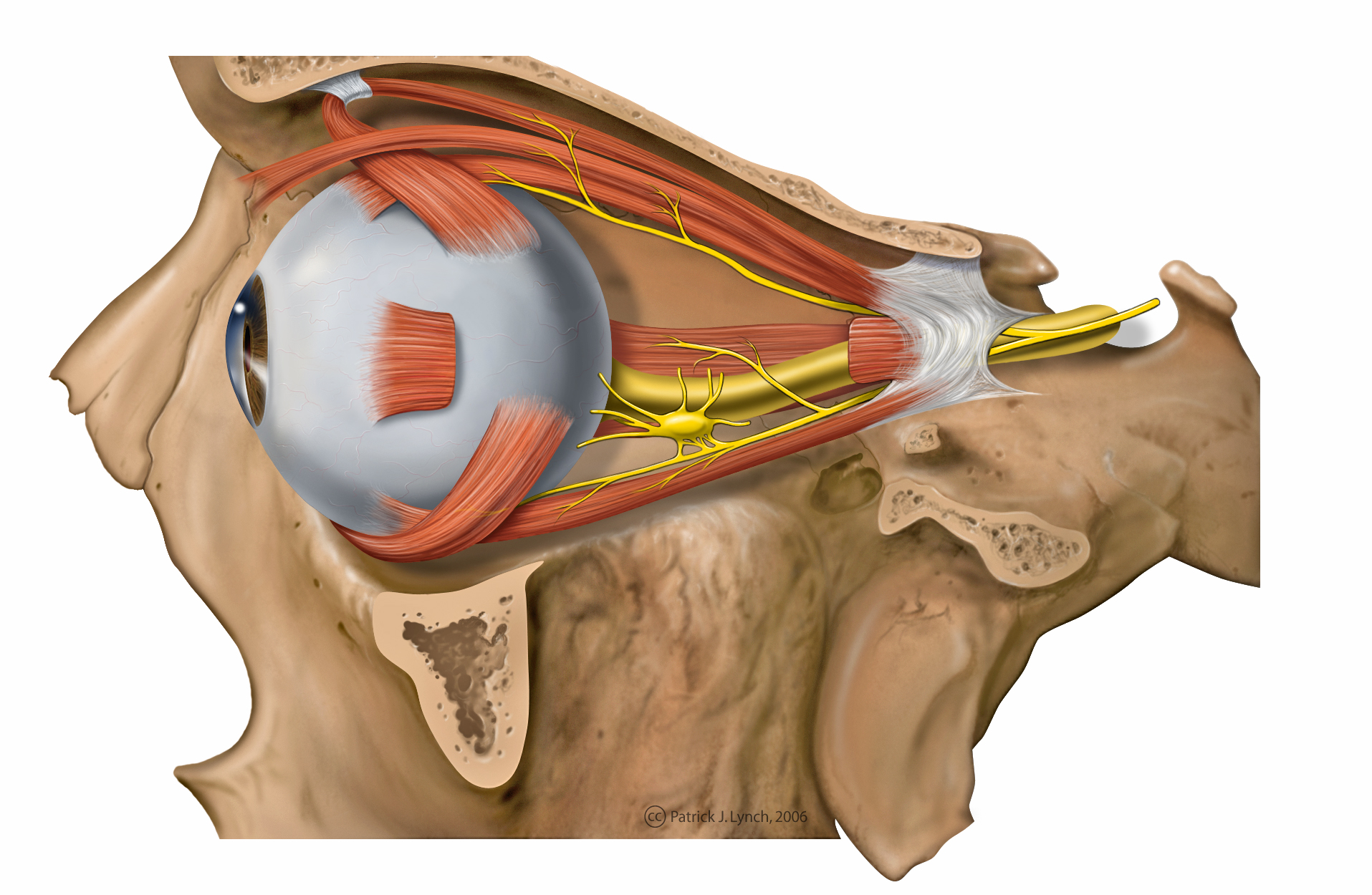
Dissection showing origins of right ocular muscles, and nerves entering by the superior orbital fissure
-
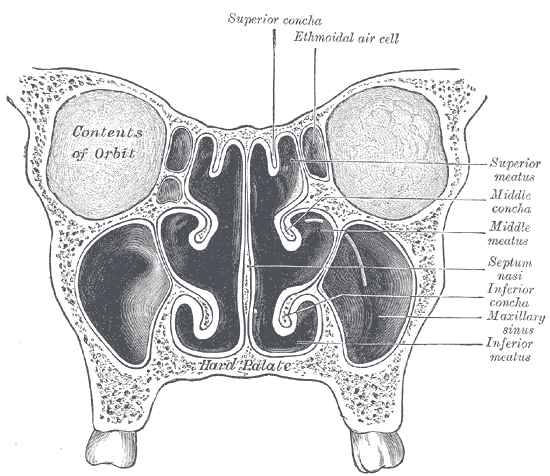
Coronal section of nasal cavities
-
-

Lateral orbit anatomy
-
Lateral orbit nerves
-

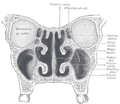
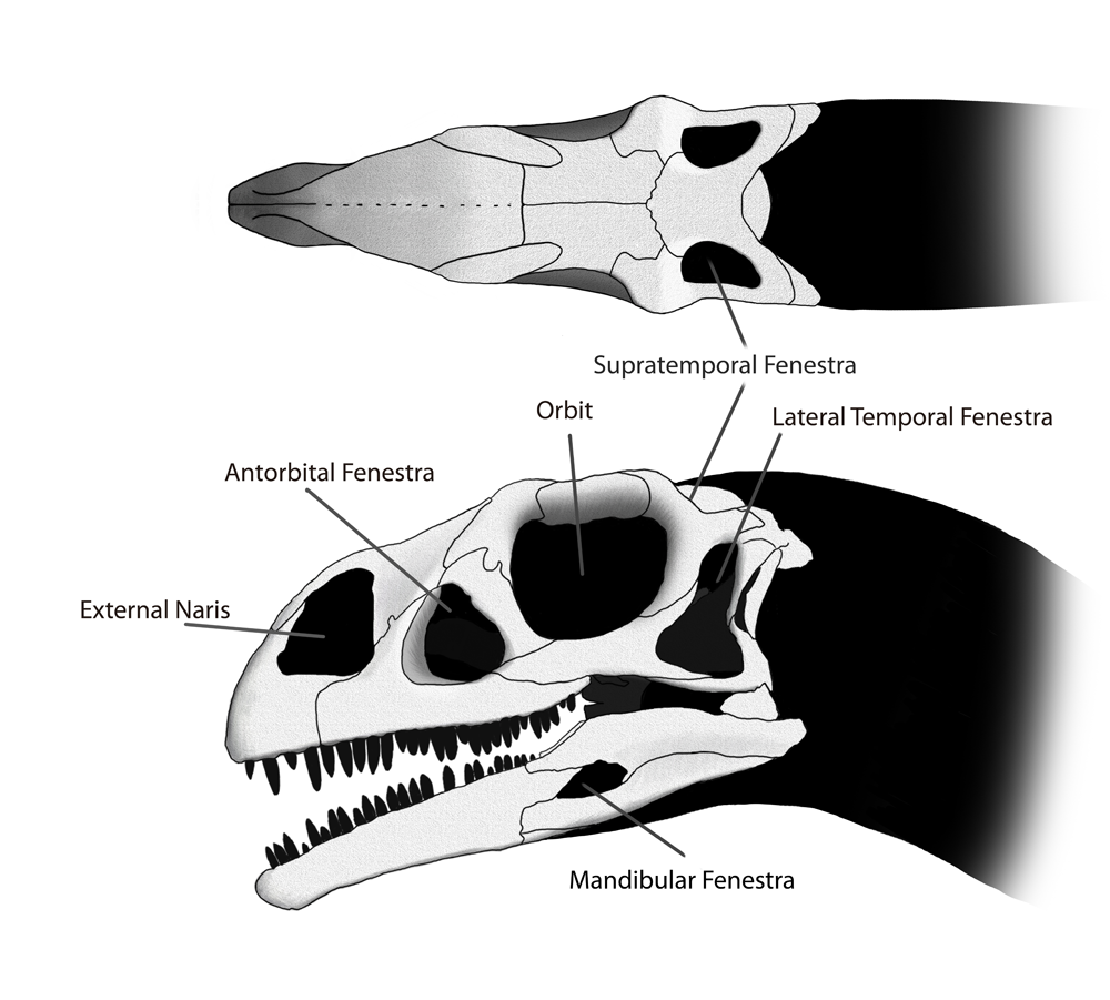
The orbit in relation to the other skull openings in the dinosaur Massospondylus
-

Orbit
-

Orbital cavity
References
- ^ "Orbit - Definition and More from the Free Merriam-Webster Dictionary". http://www.merriam-webster.com/dictionary/orbit. Retrieved 2010-03-26.
- ^ MeSH Orbit
- ^ Duane's Ophthalmology, Chapter 32 Embryology and Anatomy of the Orbit and Lacrimal System. (eds Tasman W, Jaeger EA) Lippincott/Williams & Wilkins, 2007
- ^ "eye, human."Encyclopædia Britannica from Encyclopædia Britannica 2006 Ultimate Reference Suite DVD 2009
- ^ "eye, human
- ^ Kumar V, Abbas AK, Fausto N. Robbins and Cotran Pathologic Basis of Disease. Seventh Edition. Philadephia: Elsevier Saunders, 2005, p. 1423.
- ^ Hatton MP, Rubin PA. The pathophysiology of thyroid-associated ophthalmopathy. Ophthalmol Clin North Am 15:113-119, 2002.
External links
- oph/2 at eMedicine – "Arterial Supply, Orbit"
- SUNY Labs 29:os-0501
- Atlas of anatomy at UMich eye_5
- Atlas of anatomy at UMich rsa2p4
- Interactive tutorial at anatome.ncl.ac.uk
Categories:


Wikimedia Foundation. 2010.