- Foramen lacerum
Infobox Anatomy
Name = PAGENAME
Latin =
GraySubject = 47
GrayPage = 192
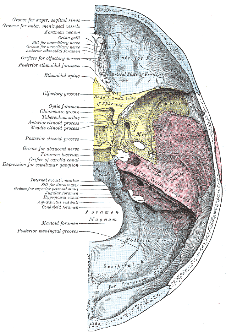
Caption = Base of the skull. Upper surface. (Foramen lacerum is labeled at center left, and is visible as the large hole between yellow sphenoid, red temporal, and blue occipital)
System =
MeshName =
MeshNumber =
DorlandsPre = f_12
DorlandsSuf = 12373219
The foramen lacerum (Latin for "lacerated piercing") is a triangular hole in the base of theskull located at the base of themedial pterygoid plate .Transit through the foramen lacerum
Several anatomy texts incorrectly state that the
internal carotid artery passes through the foramen lacerum. However, "in vivo" the foramen is actually occluded bycartilage , preventing the artery from passing through. Rather, the internal carotid artery enters the base of theskull through thecarotid canal , and travels superiorly to the cartilage occluding the foramen lacerum.However, some nerves, arteries, and veins do pass through the
cartilage plug of the foramen lacerum: theartery of pterygoid canal , thenerve of pterygoid canal , and some venous drainage.* The nerve of pterygoid canal comprises the
deep petrosal nerve and thegreater petrosal nerve the former carrying sympathetic fibres and the latterparasympathetic fibres of theautonomic nervous system toblood vessels ,mucous membranes ,salivary glands , andlacrimal glands .* Furthermore, one of the terminal branches of the
ascending pharyngeal artery (itself a branch of theexternal carotid artery ) passes through the foramen lacerum. This is one of three possible "meningeal branches " of this vessel, the ascending pharyngeal artery.* Lastly, some
emissary veins pass through the foramen lacerum. These connect the extracranialpterygoid plexus with the intracranialcavernous sinus and present an unopposed route forinfection .External links
* - "Internal view of skull."
*
* (NormanAnatomyFig|VII)
*
*
* [http://spinwarp.ucsd.edu/NeuroWeb/Anatomy/tmp-1.html Image at ucsd.edu]
Wikimedia Foundation. 2010.