- Medial palpebral ligament
-
Medial palpebral ligament 
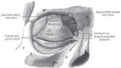
The tarsi and their ligaments. Right eye; front view. Latin l. palpebrale mediale Gray's subject #106 381 The medial palpebral ligament (tendo oculi), about 4 mm. in length and 2 mm. in breadth, is attached to the frontal process of the maxilla in front of the lacrimal groove.
Crossing the lacrimal sac, it divides into two parts, upper and lower, each attached to the medial end of the corresponding tarsus.
As the ligament crosses the lacrimal sac, a strong aponeurotic lamina is given off from its posterior surface; this expands over the sac, and is attached to the posterior lacrimal crest.
See also
This article was originally based on an entry from a public domain edition of Gray's Anatomy. As such, some of the information contained within it may be outdated.
Head and neck anatomy – accessory visual structures (TA 15.2.7, TH H3.11.08.6, GA 10.1021) Eyelid Tarsus (Meibomian pelicle) • Medial palpebral ligament • Epicanthic fold • Meibomian gland • Ciliary glands • Eyelash
Gland of ZeisLacrimal apparatus Lacrimal lake • Lacrimal gland • Lacrimal canaliculi • Lacrimal punctum • Lacrimal papilla • Nasolacrimal duct • Lacrimal sac • Lacrimal caruncle • Krause's glandsOther Periorbita • Orbital septum • Tenon's capsule • Suspensory ligament of eyeball
Conjunctiva (Plica semilunaris)
Extraocular muscles (Trochlea of superior oblique)M: EYE
anat(g/a/p)/phys/devp/prot
noco/cong/tumr, epon
proc, drug(S1A/1E/1F/1L)
Categories:- Eye anatomy
- Ligaments of the head and neck
- Ligament stubs
Wikimedia Foundation. 2010.
