- Tenon's capsule
Infobox Anatomy
Name = PAGENAME
Latin =
GraySubject = 227
GrayPage = 1024
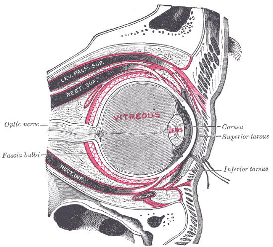
Caption = The right eye in sagittal section, showing the fascia bulbi (semidiagrammatic).
Caption2 =
System =
Precursor =
MeshName =
MeshNumber =
DorlandsPre = t_04
DorlandsSuf = 12794346The fascia bulbi (also known as the capsule of Ténon and the bulbar sheath) is a thin membrane which envelops the eyeball from the
optic nerve to the limbus, separating it from the orbital fat and forming a socket in which it plays.Its inner surface is smooth, and is separated from the outer surface of the
sclera by theperiscleral lymph space .This lymph space is continuous with the subdural and
subarachnoid cavities, and is traversed by delicate bands of connective tissue which extend between the fascia and the sclera.The fascia is perforated behind by the ciliary vessels and nerves, and fuses with the sheath of the optic nerve and with the sclera around the entrance of the
optic nerve .In front it blends with the
conjunctiva , and with it is attached to the ciliary region of the eyeball.The structure was named after
Jacques-René Tenon (1724-1816), a French surgeon and pathologist.Relations to extraocular muscles
It is perforated by the tendons of the ocular muscles, and is reflected backward on each as a tubular sheath.
The sheath of the
Obliquus superior is carried as far as the fibrous pulley of that muscle; that on theObliquus inferior reaches as far as the floor of the orbit, to which it gives off a slip.The sheaths on the recti are gradually lost in the
perimysium , but they give off important expansions.The expansion from the
Rectus superior blends with the tendon of theLevator palpebrae ; that of theRectus inferior is attached to theinferior tarsus .The expansions from the sheaths of the Recti lateralis and medialis are strong, especially that from the latter muscle, and are attached to the lacrimal and
zygomatic bone s respectively.As they probably check the actions of these two Recti they have been named the medial and lateral check ligaments.
Charles Barrett Lockwood described a thickening of the lower part of the fascia bulbi, which he named the 'suspensory ligament of the eye '. It is slung like a hammock below the eyeball, being expanded in the center, and narrow at its extremities which are attached to the zygomatic andlacrimal bone s respectively.References
Wikimedia Foundation. 2010.