- Canthus
-
For other uses, see Canthus (disambiguation).
Canthus 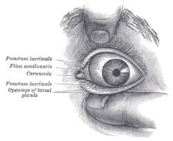
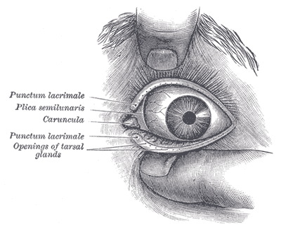
Front of left eye with eyelids separated to show medial canthus. Canthus (pl. canthi, palpebral commissures) is either corner of the eye where the upper and lower eyelids meet.[1] More specifically, the medial and lateral canthi would be described as the medial and lateral ends/angles of the palpebral fissure.
The bicanthal plane is the transversal plane linking both canthi and defines the upper boundary of the midface.
Contents
Commissures
- The lateral palpebral commissure (commissura palpebrarum lateralis; external canthus) is more acute than the medial, and the eyelids here lie in close contact with the bulb of the eye.
- The medial palpebral commissure (commissura palpebrarum medialis; internal canthus) is prolonged for a short distance toward the nose, and the two eyelids are separated by a triangular space, the lacus lacrimalis.
Surgery
Canthoplasty refers to a plastic surgery of the medial and/or lateral canthus.
A canthotomy involves cutting the canthus, often performed to release excessive orbital pressure (i.e., from orbital hemorrhage or infection).
Pathology
"Dystopia canthorum" is associated with Waardenburg syndrome.[2]
See also
References
External links
Head and neck anatomy – accessory visual structures (TA 15.2.7, TH H3.11.08.6, GA 10.1021) Eyelid Tarsus (Meibomian pelicle) • Medial palpebral ligament • Epicanthic fold • Meibomian gland • Ciliary glands • Eyelash
Palpebral fissure • Canthus
Gland of ZeisLacrimal apparatus Lacrimal lake • Lacrimal gland • Lacrimal canaliculi • Lacrimal punctum • Lacrimal papilla • Nasolacrimal duct • Lacrimal sac • Lacrimal caruncle • Krause's glandsOther Periorbita • Orbital septum • Tenon's capsule • Suspensory ligament of eyeball
Conjunctiva (Plica semilunaris)
Extraocular muscles (Trochlea of superior oblique)M: EYE
anat(g/a/p)/phys/devp/prot
noco/cong/tumr, epon
proc, drug(S1A/1E/1F/1L)
Categories:- Eye anatomy
- Eye stubs
Wikimedia Foundation. 2010.