- Lacrimal sac
-
Lacrimal sac 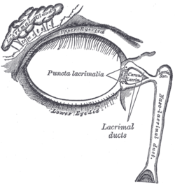
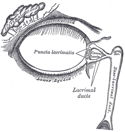
The lacrimal apparatus. Right side. (Lacrimal sac visible at upper right.) 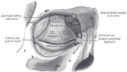
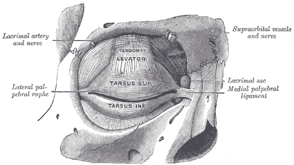
The tarsi and their ligaments. Right eye; front view. (Lacrimal sac visible at middle right.) Latin saccus lacrimalis Gray's subject #227 1028 Artery angular artery The lacrimal sac is the upper dilated end of the nasolacrimal duct, and is lodged in a deep groove formed by the lacrimal bone and frontal process of the maxilla. It connects the lacrimal canaliculi, which drain tears from the eye's surface, and the nasolacrimal duct, which conveys this fluid into the nasal cavity.
This is mainly for high amounts of tears, in which the lacrimal sac pumps inward and outward driven by the orbicularis muscle during blinking.
It is oval in form and measures from 12 to 15 mm. in length; its upper end is closed and rounded; its lower is continued into the nasolacrimal duct.
Its superficial surface is covered by a fibrous expansion derived from the medial palpebral ligament, and its deep surface is crossed by the lacrimal part of the Orbicularis oculi, which is attached to the crest on the lacrimal bone.
Like the nasolacrimal duct, the sac is lined by stratified columnar epithelium with mucus-secreting goblet cells, with surrounding connective tissue. The Lacrimal Sac also drains the eye of any debris, bacteria, dirt, etc.
Additional images
-
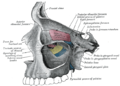
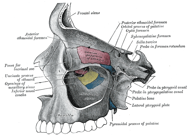
Medial wall of left orbit.
-
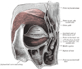
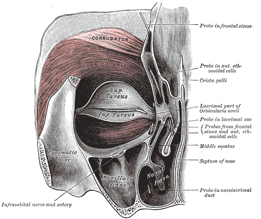
Left orbicularis oculi, seen from behind.
See also
This article was originally based on an entry from a public domain edition of Gray's Anatomy. As such, some of the information contained within it may be outdated.
Head and neck anatomy – accessory visual structures (TA 15.2.7, TH H3.11.08.6, GA 10.1021) Eyelid Tarsus (Meibomian pelicle) • Medial palpebral ligament • Epicanthic fold • Meibomian gland • Ciliary glands • Eyelash
Gland of ZeisLacrimal apparatus Lacrimal lake • Lacrimal gland • Lacrimal canaliculi • Lacrimal punctum • Lacrimal papilla • Nasolacrimal duct • Lacrimal sac • Lacrimal caruncle • Krause's glandsOther Periorbita • Orbital septum • Tenon's capsule • Suspensory ligament of eyeball
Conjunctiva (Plica semilunaris)
Extraocular muscles (Trochlea of superior oblique)M: EYE
anat(g/a/p)/phys/devp/prot
noco/cong/tumr, epon
proc, drug(S1A/1E/1F/1L)
Categories:- Eye anatomy
- Eye stubs
-
Wikimedia Foundation. 2010.