- Zonule of Zinn
-
Zonule of Zinn 
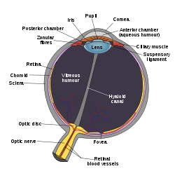
Schematic diagram of the human eye. (Zonular fibers labeled at upper left.) 
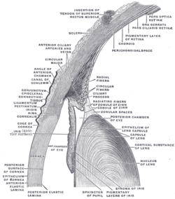
The upper half of a sagittal section through the front of the eyeball. (Zonule of Zinn visible near center.) Latin zonula ciliaris Gray's subject #226 1018 The zonule of Zinn (Zinn's membrane, ciliary zonule) (after Johann Gottfried Zinn) is a ring of fibrous strands connecting the ciliary body with the crystalline lens of the eye.
The zonule of Zinn is split into two layers: a thin layer, which lines the hyaloid fossa, and a thicker layer, which is a collection of zonular fibers. Together, the fibers are known as the suspensory ligament of the lens.[1]
Notes
References
External links
- Diagram at unmc.edu
- Diagram at eye-surgery-uk.com
- Diagram and overview at webschoolsolutions.com
- ciliary+zonule at eMedicine Dictionary
- Histology at BU 08011loa
This article was originally based on an entry from a public domain edition of Gray's Anatomy. As such, some of the information contained within it may be outdated.
Sensory system – visual system – globe of eye (TA A15.2.1–6, TH 3.11.08.0-5, GA 10.1005) Fibrous tunic (outer) Uvea/vascular tunic (middle) Retina (inner) LayersCellsPhotoreceptor cells (Cone cell, Rod cell) → (Horizontal cell) → Bipolar cell → (Amacrine cell) → Retina ganglion cell (Midget cell, Parasol cell, Bistratified cell, Giant retina ganglion cells, Photosensitive ganglion cell) → Diencephalon: P cell, M cell, K cell
Muller gliaOtherAnterior segment Posterior segment Other M: EYE
anat(g/a/p)/phys/devp/prot
noco/cong/tumr, epon
proc, drug(S1A/1E/1F/1L)
Categories:- Eye anatomy
- Ligaments
Wikimedia Foundation. 2010.


