- Optic cup (anatomical)
-
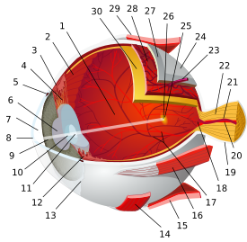
The optic cup is the white, cup-like area in the center of the optic disc.[1]
The ratio of the size of the optic cup to the optic disc (or cup-to-disc ratio) is measured to diagnose glaucoma.
References
- ^ Algazi VR, Keltner JL, Johnson CA (December 1985). "Computer analysis of the optic cup in glaucoma". Invest. Ophthalmol. Vis. Sci. 26 (12): 1759–70. PMID 4066212. http://www.iovs.org/cgi/pmidlookup?view=long&pmid=4066212.
Sensory system – visual system – globe of eye (TA A15.2.1–6, TH 3.11.08.0-5, GA 10.1005) Fibrous tunic (outer) Episcleral layer • Schlemm's canal • Trabecular meshworkUvea/vascular tunic (middle) Retina (inner) LayersCellsPhotoreceptor cells (Cone cell, Rod cell) → (Horizontal cell) → Bipolar cell → (Amacrine cell) → Retina ganglion cell (Midget cell, Parasol cell, Bistratified cell, Giant retina ganglion cells, Photosensitive ganglion cell) → Diencephalon: P cell, M cell, K cell
Muller gliaOtherMacula (Foveola, Fovea centralis) • Optic disc (Optic cup)Anterior segment Posterior segment Other M: EYE
anat(g/a/p)/phys/devp/prot
noco/cong/tumr, epon
proc, drug(S1A/1E/1F/1L)
Categories:- Eye anatomy
- Sensory organs
- Visual system
- Eye stubs
Wikimedia Foundation. 2010.


