- Cornea
-
For other uses, see Cornea (disambiguation).
Cornea 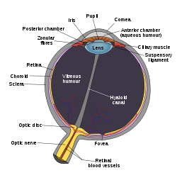
Schematic diagram of the human eye. (Cornea labeled at center top.) 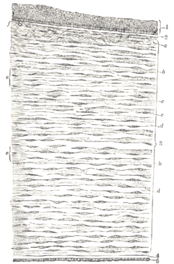
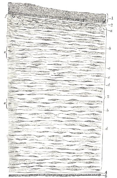
Vertical section of human cornea from near the margin. (Waldeyer.) Magnified.
1. Epithelium.
2. Anterior elastic lamina.
3. substantia propria.
4. Posterior elastic lamina.
5. Endothelium of the anterior chamber.
a. Oblique fibers in the anterior layer of the substantia propria.
b. Lamellæ the fibers of which are cut across, producing a dotted appearance.
c. Corneal corpuscles appearing fusiform in section.
d. Lamellæ the fibers of which are cut longitudinally.
e. Transition to the sclera, with more distinct fibrillation, and surmounted by a thicker epithelium.
f. Small bloodvessels cut across near the margin of the cornea.Gray's subject #225 1070 The cornea is the transparent front part of the eye that covers the iris, pupil, and anterior chamber. Together with the lens, the cornea refracts light, with the cornea accounting for approximately two-thirds of the eye's total optical power.[1][2] In humans, the refractive power of the cornea is approximately 43 dioptres.[3] While the cornea contributes most of the eye's focusing power, its focus is fixed. The curvature of the lens, on the other hand, can be adjusted to "tune" the focus depending upon the object's distance. Medical terms related to the cornea often start with the prefix "kerat-" from the Greek word κέρας, horn.
Contents
Structure
The cornea has unmyelinated nerve endings sensitive to touch, temperature and chemicals; a touch of the cornea causes an involuntary reflex to close the eyelid. Because transparency is of prime importance the cornea does not have blood vessels; it receives nutrients via diffusion from the tear fluid at the outside and the aqueous humour at the inside and also from neurotrophins supplied by nerve fibres that innervate it. In humans, the cornea has a diameter of about 11.5 mm and a thickness of 0.5–0.6 mm in the center and 0.6–0.8 mm at the periphery. Transparency, avascularity, the presence of immature resident immune cells, and immunologic privilege makes the cornea a very special tissue. The cornea has no blood supply; it gets oxygen directly through the air. Oxygen first dissolves in the tears and then diffuses throughout the cornea to keep it healthy.[4]
It borders with the sclera by the corneal limbus.
The most abundant soluble protein in mammalian cornea is albumin.[5]
In lampreys, the cornea is solely an extension of the sclera, and is separate from the skin lying above it, but in more advanced vertebrates it is always fused with the skin to form a single structure, albeit one composed of multiple layers. In fish, and aquatic vertebrates in general, the cornea plays no role in focusing light, since it has virtually the same refractive index as water.[6]
Layers
The human cornea, like those of other primates, has five layers; the corneas of cats, dogs, wolves, and other carnivores only have four.[7] From the anterior to posterior the five layers of the human cornea are:
- Corneal epithelium: a thin epithelial multicellular tissue layer (non-keratinized stratified squamous epithelium) of fast-growing and easily regenerated cells, kept moist with tears. Irregularity or edema of the corneal epithelium disrupts the smoothness of the air-tear film interface, the most significant component of the total refractive power of the eye, thereby reducing visual acuity. It is continuous with the conjunctival epithelium is composed of about 6 layers of cells which are shed constantly on the exposed layer and are regenerated by multiplication in the basal layer.
- Bowman's layer (also erroneously known as the anterior limiting membrane, when in fact it is not a membrane but a condensed layer of collagen): a tough layer that protects the corneal stroma, consisting of a similar irregularly arranged collagen fibers, mainly type I collagen fibrils, essentially a type of stroma. These fibrils interact with and attach on to each other. This layer is eight to 14 micrometres thick[8] and is absent or very thin in non-primates.[7][9]
- Corneal stroma (also substantia propria): a thick, transparent middle layer, consisting of regularly arranged collagen fibers along with sparsely distributed interconnected keratocytes, which are the cells for general repair and maintenance.[8] They are parallel and are superimposed like book pages The corneal stroma consists of approximately 200 layers of mainly type I collagen fibrils. Each layer is 1.5-2.5μm. Up to 90% of the corneal thickness is composed of stroma.[8] There are 2 theories of how transparency in the cornea comes about:
- The lattice arrangements of the collagen fibrils in the stroma. The light scatter by individual fibrils is cancelled by destructive interference from the scattered light from other individual fibrils.(Maurice, 1957)
- The spacing of the neighboring collagen fibrils in the stroma must be < 200 nm for there to be transparency. (Goldman and Benedek)
- Descemet's membrane (also posterior limiting membrane): a thin acellular layer that serves as the modified basement membrane of the corneal endothelium, from which the cells are derived. This layer is composed mainly of collagen type IV fibrils, less rigid than collagen type I fibrils, and is around 5-20μm thick, depending on the subject's age.
- Corneal endothelium: a simple squamous or low cuboidal monolayer, approx 5μm thick, of mitochondria-rich cells. These cells are responsible for regulating fluid and solute transport between the aqueous and corneal stromal compartments. (The term endothelium is a misnomer here. The corneal endothelium is bathed by aqueous humor, not by blood or lymph, and has a very different origin, function, and appearance from vascular endothelia.) Unlike the corneal epithelium the cells of the endothelium do not regenerate. Instead, they stretch to compensate for dead cells which reduces the overall cell density of the endothelium and has an impact on fluid regulation. If the endothelium can no longer maintain a proper fluid balance, stromal swelling due to excess fluids and subsequent loss of transparency will occur and this may cause corneal edema and interference with the transparency of the cornea and thus impairing the image formed.
The mnemonic "EBSDEin", read as "Ebstein" can be used to remember the layers in sequence.[10]
Another Mnemonic is "ABCDE": Anterior epithelium, Bowman's layer, Corneal stroma, Descemet's membrane and Endothelium
Keeping the cornea transparent
Upon death or removal of an eye the cornea absorbs the aqueous humor, thickens, and becomes hazy. Transparency can be restored by putting it in a warm, well-ventilated chamber at 31 °C (88 °F, the normal temperature), allowing the fluid to leave the cornea and become transparent. The cornea takes in fluid from the aqueous humor and the small blood vessels of the limbus, but a pump ejects the fluid immediately upon entry. When energy is deficient the pump may fail, or works too slowly to compensate, causing swelling. This could arise at death, but a dead eye can be placed in a warm chamber and the reservoirs of sugar and glycogen can keep the cornea transparent for at least 24 hours. The endothelium controls this pumping action, and as discussed above, damage thereof is more serious, and is a cause of opaqueness and swelling. When damage to the cornea occurs, such as in a viral infection, the collagen used to repair the process is not regularly arranged, leading to an opaque patch (leukoma). When a cornea is needed for transplant, as from an eye bank, the best procedure is to remove the cornea from the eyeball, preventing the cornea from absorbing the aqueous humor.[8]
Innervation
The cornea is one of the most sensitive tissues of the body, as it is densely innervated with sensory nerve fibres via the ophthalmic division of the trigeminal nerve by way of 70–80 long ciliary nerves and short ciliary nerves. The ciliary nerves run under the endothelium and exit the eye through holes in the sclera apart from the optic nerve (which transmits only optic signals).[8]
The nerves enter the cornea via three levels; scleral, episcleral and conjunctival. Most of the bundles give rise by subdivision to a network in the stroma, from which fibres supply the different regions. The three networks are midstromal, subepithelial/Bowman's layer, and epithelium. The receptive fields of each nerve ending are very large, and may overlap.
Corneal nerves of the subepithelial layer terminate near superficial epithelial layer of the cornea in a logarithmic spiral pattern.[11] The density of epithelial nerves decreases with age, especially after the seventh decade.[12]
Refractive nature
The optical component is concerned with producing a reduced inverted image on the retina. The eye's optical system consists of not only two but four surfaces—two on the cornea, two on the lens. Rays are refracted toward the midline. Distant rays, due to their parallel nature, converge to a point on the retina. The cornea admits light at the greatest angle. The aqueous and vitreous humors both have a refractive index of 1.336, whereas the cornea has a refractive index of 1.376. Because the change in refractive index between cornea and aqueous humor is relatively small compared to the change at the air–cornea interface, it has a negligible refractive effect, typically -6 diopters.[8]
Diseases and disorders
Main article: Eye diseaseTreatment and management
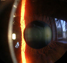 Slit lamp image of the cornea, iris and lens
Surgical procedures
Various refractive eye surgery techniques change the shape of the cornea in order to reduce the need for corrective lenses or otherwise improve the refractive state of the eye. In many of the techniques used today, reshaping of the cornea is performed by photoablation using the excimer laser.
If the corneal stroma develops visually significant opacity, irregularity, or edema, a cornea of a deceased donor can be transplanted. Because there are no blood vessels in the cornea, there are also few problems with rejection of the new cornea.
There are also synthetic corneas (keratoprostheses) in development. Most are merely plastic inserts, but there are also those composed of biocompatible synthetic materials that encourage tissue ingrowth into the synthetic cornea, thereby promoting biointegration.
Non-surgical procedures
Orthokeratology is a method using specialized hard or rigid gas-permeable contact lenses to transiently reshape the cornea in order to improve the refractive state of the eye or reduce the need for eyeglasses and contact lenses.
In 2009, researchers at the University of Pittsburgh Medical center demonstrated that stem cells collected from human corneas can restore transparency without provoking a rejection response in mice with corneal damage.[13]
See also
References
Notes
- ^ Cassin, B. and Solomon, S. Dictionary of Eye Terminology. Gainsville, Florida: Triad Publishing Company, 1990.
- ^ Goldstein, E. Bruce. Sensation & Perception. 7th Edition. Canada: Thompson Wadsworth, 2007.
- ^ Najjar, Dany "Clinical optics and refraction"
- ^ The Association of Contact Lens Manufacturers "Why does the cornea need oxygen?"
- ^ Nees DW, Fariss RN, Piatigorsky J (August 2003). "Serum albumin in mammalian cornea: implications for clinical application". Invest. Ophthalmol. Vis. Sci. 44 (8): 3339–45. doi:10.1167/iovs.02-1161. PMID 12882779. http://www.iovs.org/cgi/pmidlookup?view=long&pmid=12882779.
- ^ Romer, Alfred Sherwood; Parsons, Thomas S. (1977). The Vertebrate Body. Philadelphia, PA: Holt-Saunders International. pp. 461–462. ISBN 0-03-910284-X.
- ^ a b Merindano MD; Costa J; Canals M; Potau JM, and Ruano D. "A comparative study of Bowman's layer in some mammals: Relationships with other constituent corneal structures." European Journal of Anatomy. Volume 6, Number 3, December 2002.
- ^ a b c d e f "eye, human."Encyclopædia Britannica from Encyclopædia Britannica 2006 Ultimate Reference Suite DVD 2009
- ^ Hayashi, S; Osawa, T; Tohyama, K (2002). "Comparative observations on corneas, with special reference to Bowman's layer and Descemet's membrane in mammals and amphibians". Journal of morphology 254 (3): 247–58. doi:10.1002/jmor.10030. PMID 12386895.
- ^ [1] Layers of cornea: Mnemonic
- ^ Yu CQ, Rosenblatt MI. Transgenic corneal neurofluorescence in mice: a new model for in vivo investigation of nerve structure and regeneration. Invest Ophthalmol Vis Sci. 2007 Apr;48(4):1535-42.
- ^ "Mapping the entire human corneal nerve architecture". Exp Eye Res. 2010 Oct;91(4):513-23. 2010 Oct;91(4):513-23. PMID 20650270. http://www.ncbi.nlm.nih.gov/pubmed/20650270?dopt=Abstract. Retrieved 2011-03-10-2011.
- ^ "Stem Cell Therapy Makes Cloudy Corneas Clear, According To Pitt Researchers". Medical News Today. 13 April 2009. http://www.medicalnewstoday.com/articles/145528.php. Retrieved 2009-06-04.
General references
- Daxer A, Misof K, Grabner B, Ettl A, Fratzl P. "Collagen fibrils in the human corneal stroma: structure and aging." Invest Ophthalmol Vis Sci. 1998 Mar;39(3):644-8. PMID 9501878.
- Daxer A, Fratzl P. "Collagen fibril orientation in the human corneal stroma and its implication in keratoconus." Invest Ophthalmol Vis Sci. 1997 Jan;38(1):121-9. PMID 9008637.
- Fratzl P, Daxer A. "Structural transformation of collagen fibrils in corneal stroma during drying. An x-ray scattering study." Biophys J. 1993 Apr;64(4):1210-4. PMID 8494978.
External links
- Atlas of anatomy at UMich eye_1 - "Sagittal Section Through the Eyeball"
- Facts About The Cornea and Corneal Disease National Eye Institute (NEI)
- Keratoconus (Irregular Astigmatism), Patient Support
- Keratos, Patient Support
- Corneal Endothelial pump
Sensory system – visual system – globe of eye (TA A15.2.1–6, TH 3.11.08.0-5, GA 10.1005) Fibrous tunic (outer) Episcleral layer • Schlemm's canal • Trabecular meshworkCornea

Uvea/vascular tunic (middle) Retina (inner) LayersCellsPhotoreceptor cells (Cone cell, Rod cell) → (Horizontal cell) → Bipolar cell → (Amacrine cell) → Retina ganglion cell (Midget cell, Parasol cell, Bistratified cell, Giant retina ganglion cells, Photosensitive ganglion cell) → Diencephalon: P cell, M cell, K cell
Muller gliaOtherAnterior segment Posterior segment Other M: EYE
anat(g/a/p)/phys/devp/prot
noco/cong/tumr, epon
proc, drug(S1A/1E/1F/1L)
Categories:- Eye anatomy


Wikimedia Foundation. 2010.