- Iris dilator muscle
-
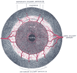
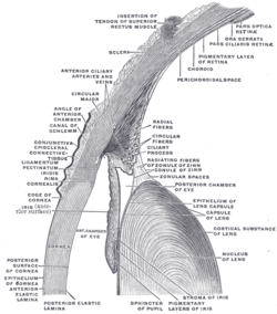
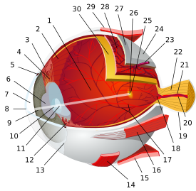
Iris dilator muscle Iris, front view. (Muscle visible but not labeled.) The upper half of a sagittal section through the front of the eyeball. (Iris dilator muscle is NOT labeled and not to be confused with "Radiating fibers" labeled near center, which are part of the ciliary muscle.) Latin musculus dilatator pupillae Gray's subject #225 1013 Origin outer margins of iris[1] Insertion inner margins of iris[1] Artery Nerve Long ciliary nerves (sympathetics) Actions dilates pupil Antagonist iris sphincter muscle The iris dilator muscle (pupil dilator muscle, pupillary dilator, radial muscle of iris, radiating fibers), is a smooth muscle[2] of the eye, running radially in the iris and therefore fit as a dilator. It has its origin from the anterior epithelium.[3] It is innervated by the sympathetic system, which acts by releasing noradrenaline, which acts on α1-receptors.[4] Thus, when presented with a threatening stimuli that activates the fight-or-flight response, this innervation contracts the muscle and dilates the iris, thus temporarily letting more light reach the retina.
Contents
Additional images
See also
References
- ^ a b Gest, Thomas R; Burkel, William E. "Anatomy Tables - Eye." Medical Gross Anatomy. 2000. University of Michigan Medical School. 5 Jan. 2010 <http://anatomy.med.umich.edu/nervous_system/eye_tables.html>.
- ^ jneurosci.org Muscarinic and Nicotinic Synaptic Activation of the Developing..
- ^ "eye, human." Encyclopædia Britannica. Encyclopaedia Britannica Ultimate Reference Suite. Chicago: Encyclopædia Britannica, 2010.
- ^ Rang, H. P. (2003). Pharmacology. Edinburgh: Churchill Livingstone. ISBN 0-443-07145-4. Page 163
External links
- Description of function at tedmontgomery.com
- Slide at mscd.edu
- dilator+pupillae+muscle at eMedicine Dictionary
- Histology at BU 08010loa
Sensory system – visual system – globe of eye (TA A15.2.1–6, TH 3.11.08.0-5, GA 10.1005) Fibrous tunic (outer) Episcleral layer • Schlemm's canal • Trabecular meshworkUvea/vascular tunic (middle) Retina (inner) LayersCellsPhotoreceptor cells (Cone cell, Rod cell) → (Horizontal cell) → Bipolar cell → (Amacrine cell) → Retina ganglion cell (Midget cell, Parasol cell, Bistratified cell, Giant retina ganglion cells, Photosensitive ganglion cell) → Diencephalon: P cell, M cell, K cell
Muller gliaOtherAnterior segment Posterior segment Other M: EYE
anat(g/a/p)/phys/devp/prot
noco/cong/tumr, epon
proc, drug(S1A/1E/1F/1L)
Sensory system: Visual system and eye movement pathways Visual perception 1° (Bipolar cell of Retina) → 2° (Ganglionic cell) → 3° (Optic nerve → Optic chiasm → Optic tract → LGN of Thalamus) → 4° (Optic radiation → Cuneus and Lingual gyrus of Visual cortex → Blobs → Globs)Muscles of orbit TrackingHorizontal gazeVertical gazePupillary reflex Pupillary dilation1° (Posterior hypothalamus → Ciliospinal center) → 2° (Superior cervical ganglion) → 3° (Sympathetic root of ciliary ganglion → Nasociliary nerve → Long ciliary nerves → Iris dilator muscle)1° (Retina → Optic nerve → Optic chiasm → Optic tract → Visual cortex → Brodmann area 19 → Pretectal area) → 2° (Edinger-Westphal nucleus) → 3° (Short ciliary nerves → Ciliary ganglion → Ciliary muscle)Circadian rhythm M: EYE
anat(g/a/p)/phys/devp/prot
noco/cong/tumr, epon
proc, drug(S1A/1E/1F/1L)
Categories:- Muscular system
- Eye anatomy
- Muscle stubs
- Eye stubs
Wikimedia Foundation. 2010.


