- Vergence
-
A vergence is the simultaneous movement of both eyes in opposite directions to obtain or maintain single binocular vision.[1].
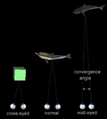
When a creature with binocular vision looks at an object, the eyes must rotate around a vertical axis so that the projection of the image is in the centre of the retina in both eyes. To look at an object closer by, the eyes rotate towards each other (convergence), while for an object farther away they rotate away from each other (divergence). Exaggerated convergence is called cross eyed viewing (focussing on the nose for example) . When looking into the distance, the eyes diverge until parallel, effectively fixating the same point at infinity (or very far away).
Vergence movements are closely connected to accommodation of the eye. Under normal conditions, changing the focus of the eyes to look at an object at a different distance will automatically cause vergence and accommodation.
As opposed to the 500°/s velocity of saccade movements, vergence movements are far slower, around 25°/s. The extraocular muscles may have two types of fiber each with its own nerve supply, hence a dual mechanism.[citation needed]
Vergence dysfunction
A number of vergence dysfunctions exist:[2][3]
- Basic exophoria
- Convergence insufficiency
- Divergence excess
- Basic esophoria
- Convergence excess
- Divergence insufficiency
- Fusional vergence dysfunction
- Vertical phorias
See also
References
- ^ Cassin, B. Dictionary of Eye Terminology. Solomon S.. Gainesville, Fl: Triad Publishing Company. ISBN 0937404683.
- ^ American Optometric Association. Optometric Clinical Practice Guideline: Care of the Patient with Accommodative and Vergence Dysfunction. 1998.
- ^ Duane A. "A new classification of the motor anomalies of the eyes based upon physiological principles, together with their symptoms, diagnosis and treatment." Ann Ophthalmol. Otolaryngol. 5:969.1869;6:94 and 247.1867.
Sensory system: Visual system and eye movement pathways Visual perception 1° (Bipolar cell of Retina) → 2° (Ganglionic cell) → 3° (Optic nerve → Optic chiasm → Optic tract → LGN of Thalamus) → 4° (Optic radiation → Cuneus and Lingual gyrus of Visual cortex → Blobs → Globs)Muscles of orbit TrackingHorizontal gazeVertical gazePupillary reflex Pupillary dilation1° (Posterior hypothalamus → Ciliospinal center) → 2° (Superior cervical ganglion) → 3° (Sympathetic root of ciliary ganglion → Nasociliary nerve → Long ciliary nerves → Iris dilator muscle)1° (Retina → Optic nerve → Optic chiasm → Optic tract → Visual cortex → Brodmann area 19 → Pretectal area) → 2° (Edinger-Westphal nucleus) → 3° (Short ciliary nerves → Ciliary ganglion → Ciliary muscle)Circadian rhythm M: EYE
anat(g/a/p)/phys/devp/prot
noco/cong/tumr, epon
proc, drug(S1A/1E/1F/1L)
Categories:- Eye
- Eye stubs
Wikimedia Foundation. 2010.

