- Vestibulo-ocular reflex
-
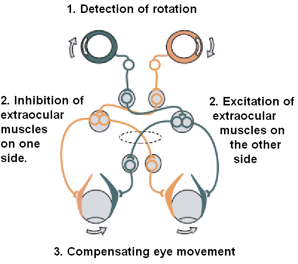
 The vestibulo-ocular reflex. A rotation of the head is detected, which triggers an inhibitory signal to the extraocular muscles on one side and an excitatory signal to the muscles on the other side. The result is a compensatory movement of the eyes.
The vestibulo-ocular reflex (VOR) is a reflex eye movement that stabilizes images on the retina during head movement by producing an eye movement in the direction opposite to head movement, thus preserving the image on the center of the visual field. For example, when the head moves to the right, the eyes move to the left, and vice versa. Since slight head movement is present all the time, the VOR is very important for stabilizing vision: patients whose VOR is impaired find it difficult to read using print, because they cannot stabilize the eyes during small head tremors. The VOR does not depend on visual input and works even in total darkness or when the eyes are closed. However, in the presence of light, the fixation reflex is also added to the movement.[1]
In lower animals, the gravity organs and eyes are strictly connected. The fish, for instance, moves its eyes by reflex when its tail is moved. Humans have semicircular canals, neck muscle "stretch" receptors, and the utricle (gravity organ). Though the semicircular canals cause most of the reflexes which are responsive to acceleration, the maintaining of balance is mediated by the stretch of neck muscles and the pull of gravity on the utricle (otolith organ) of the inner ear.[1]
The VOR has both rotational and translational aspects. When the head rotates about any axis (horizontal, vertical, or torsional) distant visual images are stabilized by rotating the eyes about the same axis, but in the opposite direction.[2] When the head translates, for example during walking, the visual fixation point is maintained by rotating gaze direction in the opposite direction, by an amount that depends on distance.[3]
Contents
Circuit
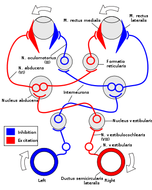
The VOR is ultimately driven by signals from the vestibular apparatus in the inner ear. The semicircular canals detect head rotation and drive the rotational VOR, whereas the otoliths detect head translation and drive the translational VOR. The main "direct path" neural circuit for the horizontal rotational VOR is fairly simple. It starts in the vestibular system, where semicircular canals get activated by head rotation and send their impulses via the vestibular nerve (cranial nerve VIII) through Scarpa's ganglion and end in the vestibular nuclei in the brainstem. From these nuclei, fibers cross to the contralateral cranial nerve VI nucleus (abducens nucleus). There they synapse with 2 additional pathways. One pathway projects directly to the lateral rectus of eye via the abducens nerve. Another nerve tract projects from the abducens nucleus by the medial longitudinal fasciculus to the oculomotor nuclei, which contain motorneurons that drive eye muscle activity, specifically activating the medial rectus muscles of the eye through the oculomotor nerve.
Another pathway (not in picture) directly projects from the vestibular nucleus through the ascending tract of Dieters to the ipsilateral medial rectus motoneuron. In addition there are inhibitory vestibular pathways to the ipsilateral abducens nucleus. However no direct vestibular neuron to medial rectus motoneuron pathway exists.[4]
Similar pathways exist for the vertical and torsional components of the VOR.
In addition to these direct pathways, which drive the velocity of eye rotation, there is an indirect pathway that builds up the position signal needed to prevent the eye from rolling back to center when the head stops moving. This pathway is particularly important when the head is moving slowly, because here position signals dominate over velocity signals. David A. Robinson discovered that the eye muscles require this dual velocity-position drive, and also proposed that it must arise in the brain by mathematically integrating the velocity signal and then sending the resulting position signal to the motoneurons. Robinson was correct: the 'neural integrator' for horizontal eye position was found in the nucleus prepositus hypoglossi[5] in the medulla, and the neural integrator for vertical and torsional eye positions was found in the interstitial nucleus of Cajal[6] in the midbrain. The same neural integrators also generate eye position for other conjugate eye movements such as saccades and smooth pursuit.
Excitatory example
For instance, if the head is turned clockwise as seen from above, then excitatory impulses are sent from the semicircular canal on the right side via the vestibular nerve (cranial nerve VIII) through Scarpa's ganglion and end in the right vestibular nuclei in the brainstem. From this nuclei excitatory fibers cross to the left abducens nucleus. There they project and stimulate the lateral rectus of the left eye via the abducens nerve. In addition, by the medial longitudinal fasciculus and oculomotor nuclei, they activate the medial rectus muscles on the right eye. As a result, both eyes will turn counterclockwise.
Furthermore, some neurons from the right vestibular nucleus directly stimulate the right medial rectus motoneurons, and inhibits the right abducens nucleus.
Speed
The vestibulo-ocular reflex needs to be fast: for clear vision, head movement must be compensated almost immediately; otherwise, vision corresponds to a photograph taken with a shaky hand. To achieve clear vision, signals from the semicircular canals are sent as directly as possible to the eye muscles: the connection involves only three neurons, and is correspondingly called the three neuron arc. Using these direct connections, eye movements lag the head movements by less than 10 ms,[7] and thus the vestibulo-ocular reflex is one of the fastest reflexes in the human body.
Gain
The "gain" of the VOR is defined as the change in the eye angle divided by the change in the head angle during the head turn. Ideally the gain of the rotational VOR is 1.0. The gain of the horizontal and vertical VOR is usually close to one, but the gain of the torsional VOR (rotation around the line of sight) is generally low.[2] The gain of the translational VOR has to be adjusted for distance, because of the geometry of motion parallax. When the head translates, the angular direction of near targets changes faster than the angular direction of far targets.[3]
If the gain of the VOR is wrong (different from 1)—for example, if eye muscles are weak, or if a person puts on a new pair of eyeglasses—then head movement results in image motion on the retina, resulting in blurred vision. Under such conditions, motor learning adjusts the gain of the VOR to produce more accurate eye motion. This is what is referred to as VOR adaptation.
Ethanol consumption can disrupt the VOR, reducing dynamic visual acuity.[8]
Testing
This reflex can be tested by the Rapid head impulse test or Halmagyi-Curthoys-test, in which the head is rapidly moved to the side with force, and is controlled if the eyes succeed to remain looking in the same direction. When the function of the right balance system is reduced, by a disease or by an accident, quick head movement to the right cannot be sensed properly any more. As a consequence, no compensatory eye movement is generated, and the patient cannot fixate a point in space during this rapid head movement.
Another way of testing the VOR response is a caloric reflex test, which is an attempt to induce nystagmus (compensatory eye movement in the absence of head motion) by pouring cold or warm water into the ear. Also available is bi-thermal air caloric irrigations, in which warm and cool air is administered into the ear.
Comatose patients
In comatose patients, once it has been determined that the cervical spine is intact, a test of the vestibulo-ocular reflex can be performed by turning the head to one side. If the brainstem is intact, the eyes will move conjugately away from the direction of turning (as if still looking at the examiner rather than fixed straight ahead). This is how a doll's eyes would move. So having "doll's eyes" is a sign that a comatose patient's brainstem is still intact.
Testing complications
Currently, vestibulo-ocular reflexes can only be comprehensively tested in specially equipped laboratories. The tests sometimes provide valuable diagnostic information; but the laboratory setting is unnatural, the tests are time-consuming, and the people being tested are often asymptomatic while in the lab. A device capable of tracking eye movement outside the laboratory would be very useful to clinicians. Steven Rauch, of the Massachusetts Eye and Ear Infirmary, is in the process of developing an ambulatory vestibular monitoring device.
Role of cerebellum
The cerebellum is essential for motor learning to correct the VOR in order to ensure accurate eye movement. Motor learning in the VOR is in many ways analogous to classical eyeblink conditioning, since the circuits are homologous and the molecular mechanisms are similar.
See also
- Caloric reflex test
- Semicircular canals
- Vestibular system
- Pursuit movement
- Image stabilization
References
- ^ a b "Sensory Reception: Human Vision: Structure and function of the Human Eye" vol. 27, p. 179 Encyclopaedia Britannica, 1987
- ^ a b Crawford JD, Vilis T. (1991) Axes of eye rotation and Listing's law during rotations of the head. J Neurophysiol. 65(3):407-23.
- ^ a b Angelaki DE. (2004) Eyes on target: what neurons must do for the vestibuloocular reflex during linear motion. J Neurophysiol. 92(1):20-35.
- ^ Straka H, Dieringer N (2004). "Basic organization principles of the VOR: lessons from frogs". Prog. Neurobiol. 73 (4): 259–309. doi:10.1016/j.pneurobio.2004.05.003. PMID 15261395.
- ^ Cannon SC, Robinson DA. (1987) Loss of the neural integrator of the oculomotor system from brain stem lesions in monkey. J Neurophysiol. 1987 57(5):1383-409.
- ^ Crawford JD, Cadera W, Vilis T. (1991) Generation of torsional and vertical eye position signals by the interstitial nucleus of Cajal. Science. 252(5012):1551-3.
- ^ Aw, S. T., G. M. Halmagyi, T. Haslwanter, I. S. Curthoys, R. A. Yavor and M. J. Todd (1996). "Three-dimensional vector analysis of the human vestibuloocular reflex in response to high-acceleration head rotations. II. responses in subjects with unilateral vestibular loss and selective semicircular canal occlusion." J Neurophysiol 76(6): 4021-30.
- ^ "Effect of Ethanol on visual-vestibular interactions during vertical linear body acceleration". http://cat.inist.fr/?aModele=afficheN&cpsidt=15155766.
Nervous system physiology: neurophysiology - reflex Cranial nerve midbrain: Pupillary light reflex · Accommodation reflex
pons/medulla: Jaw jerk reflex · Corneal reflex · Caloric reflex test/Vestibulo-ocular reflex/Oculocephalic reflex · Pharyngeal (gag) reflexStretch reflexes upper limb: Biceps reflex C5/C6 · Brachioradialis reflex C6 · Triceps reflex C7/C8
lower limb: Patellar reflex L2-L4 · Ankle jerk reflex S1/S2 · Plantar reflex L5-S2Primitive reflexes Lists Cardiovascular Bainbridge reflex · Bezold-Jarisch reflex · Coronary reflex · Mammalian diving reflex · Oculocardiac reflexReflex bradycardia · Reflex tachycardiaRespiratoryChurchill-Cope reflexOther Acoustic reflex · H-reflex · Golgi tendon reflex · Optokinetic · Startle reaction · Withdrawal reflex (Crossed extensor reflex)External links
- (Video) Head Impulse Testing site (vHIT) Site with thorough information about vHIT
- Motor Learning in the VOR in Mice at edboyden.org
- Review on VOR adaptation via slides at Johns Hopkins University
- MeSH Vestibulo-Ocular+Reflex
- Center for Integration of Medicine and Innovative Technology - Testing device development
- ent/482 at eMedicine - "Vestibuloocular Reflex Testing"
Sensory system: Visual system and eye movement pathways Visual perception 1° (Bipolar cell of Retina) → 2° (Ganglionic cell) → 3° (Optic nerve → Optic chiasm → Optic tract → LGN of Thalamus) → 4° (Optic radiation → Cuneus and Lingual gyrus of Visual cortex → Blobs → Globs)Muscles of orbit TrackingHorizontal gazeVertical gazeVestibulo-ocular reflexPupillary reflex Pupillary dilation1° (Posterior hypothalamus → Ciliospinal center) → 2° (Superior cervical ganglion) → 3° (Sympathetic root of ciliary ganglion → Nasociliary nerve → Long ciliary nerves → Iris dilator muscle)1° (Retina → Optic nerve → Optic chiasm → Optic tract → Visual cortex → Brodmann area 19 → Pretectal area) → 2° (Edinger-Westphal nucleus) → 3° (Short ciliary nerves → Ciliary ganglion → Ciliary muscle)Circadian rhythm M: EYE
anat(g/a/p)/phys/devp/prot
noco/cong/tumr, epon
proc, drug(S1A/1E/1F/1L)
Categories:- Reflexes
- Vision

Wikimedia Foundation. 2010.