- Cuneus
-
Brain: Cuneus 
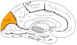
Medial surface of left cerebral hemisphere. (Cuneus visible at left as orange.) 
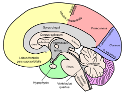
Medial view of a halved human brain Artery posterior cerebral artery NeuroNames hier-139 - Cuneus (Latin for "wedge"; plural, cunei) is also the architectural term applied to the wedge-shaped divisions of the Roman theatre separated by the scalae or stairways; see Vitruvius v. 4. This shape also occurred in medieval architecture.
The cuneus is a portion of the human brain in the occipital lobe.
The cuneus (Brodmann area 17) receives visual information from the contralateral superior retina representing the inferior visual field. It is most known for its involvement in basic visual processing. Pyramidal cells in the cuneus (striate cortex) project to extrastriate cortices (BA 18,19). The mid-level visual processing that occurs in the extrastriate projection fields of the cuneus are modulated by extraretinal effects, like attention, working memory, and reward expectation.
In addition to its traditional role as a site for basic visual processing, gray matter volume in the cuneus is associated with better inhibitory control in bipolar depression patients.[1] Pathologic gamblers have higher activity in the dorsal visual processing stream including the cuneus relative to controls.[2]
Gallery
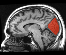
 Position of cuneus(red) of left cerebral hemisphere.
References
- ^ Haldane M, Cunningham G, Androutsos C, Frangou S (March 2008). "Structural brain correlates of response inhibition in Bipolar Disorder I". Journal of Psychopharmacology 22 (2): 138–43. doi:10.1177/0269881107082955. PMID 18308812.
- ^ Crockford DN, Goodyear B, Edwards J, Quickfall J, el-Guebaly N (November 2005). "Cue-induced brain activity in pathological gamblers". Biological Psychiatry 58 (10): 787–95. doi:10.1016/j.biopsych.2005.04.037. PMID 15993856.
Sensory system: Visual system and eye movement pathways Visual perception 1° (Bipolar cell of Retina) → 2° (Ganglionic cell) → 3° (Optic nerve → Optic chiasm → Optic tract → LGN of Thalamus) → 4° (Optic radiation → Cuneus and Lingual gyrus of Visual cortex → Blobs → Globs)Muscles of orbit TrackingHorizontal gazeVertical gazePupillary reflex Pupillary dilation1° (Posterior hypothalamus → Ciliospinal center) → 2° (Superior cervical ganglion) → 3° (Sympathetic root of ciliary ganglion → Nasociliary nerve → Long ciliary nerves → Iris dilator muscle)1° (Retina → Optic nerve → Optic chiasm → Optic tract → Visual cortex → Brodmann area 19 → Pretectal area) → 2° (Edinger-Westphal nucleus) → 3° (Short ciliary nerves → Ciliary ganglion → Ciliary muscle)Circadian rhythm M: EYE
anat(g/a/p)/phys/devp/prot
noco/cong/tumr, epon
proc, drug(S1A/1E/1F/1L)
 This article incorporates text from a publication now in the public domain: Chisholm, Hugh, ed (1911). Encyclopædia Britannica (11th ed.). Cambridge University Press.Categories:
- Nervous system
- Neuroanatomy
- Architectural elements
- Neuroanatomy stubs
- Architectural element stubs
Wikimedia Foundation. 2010.

