- Optic chiasm
-
Brain: Optic chiasm 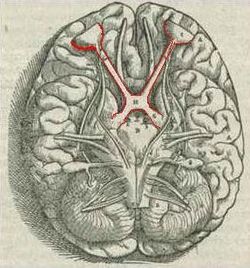
Visual pathway with optic chiasm (X shape outlined, red) (1543 image from Andreas Vesalius' Fabrica) Latin chiasma opticum Gray's subject #197 883 MeSH Optic+chiasm NeuroLex ID birnlex_1416 The optic chiasm or optic chiasma (Greek χίασμα, "crossing", from the Greek χιαζω 'to mark with an X', after the Greek letter 'Χ', chi) is the part of the brain where the optic nerves (CN II) partially cross. The optic chiasm is located at the bottom of the brain immediately below the hypothalamus.[1]
Contents
Pathways
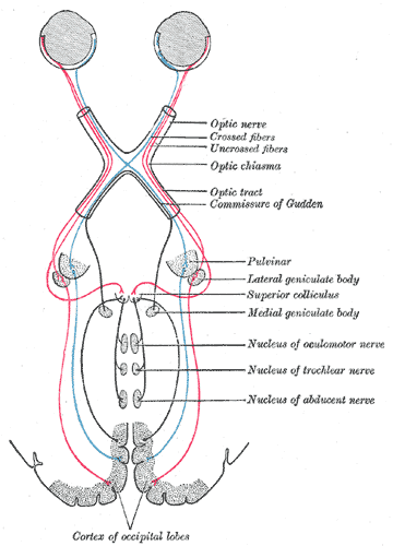
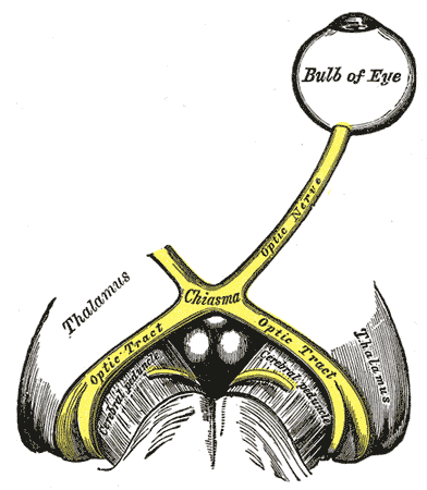
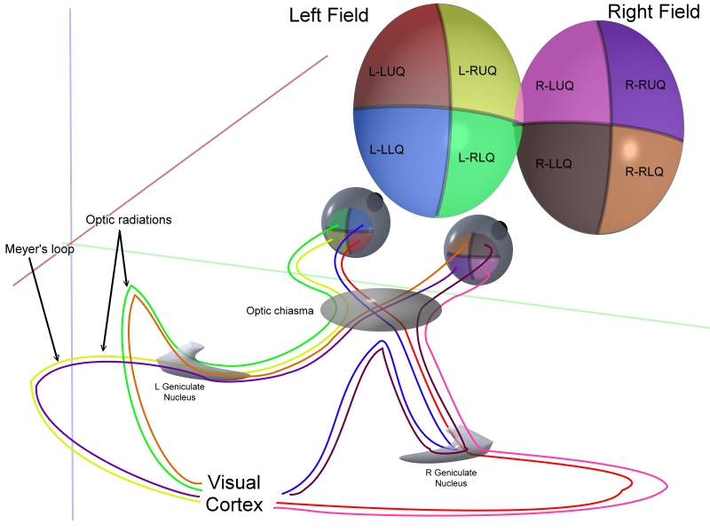
The images on the nasal sides of each retina cross over to the opposite side of the brain via the optic nerve at the optic chiasm. The temporal images, on the other hand, stay on the same side. This allows the images from either side of the field from both eyes to be transmitted to the appropriate side of the brain, combining the sides together. Beyond the optic chiasm, with crossed and uncrossed fibers, optic nerves become optic tracts. This allows for parts of both eyes that attend to the right visual field to be processed in the left visual system in the brain, and vice versa. This is linked to skin sensation which reaches the opposite side of the body, after reaching the diencephalon (rear forebrain). Decussation is an adaptive feature of frontally oriented eyes and therefore having binocular vision. Some animals, with laterally positioned eyes, have little binocular vision, so there is a more complete crossover of visual signals. The signals are passed on to the lateral geniculate body, in turn giving them to the occipital cortex (the outer matter of the rear brain).[2]
Optic chiasm in cats
In Siamese cats with certain genotypes of the albino gene, this wiring is disrupted, with less of the nerve-crossing than is normal, as a number of scholars have reported.[3] To compensate for lack of crossing in their brains, they cross their eyes (strabismus).[4]
This is also seen in albino tigers, as Guillery & Kaas report.[5]
Additional images
-
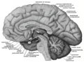
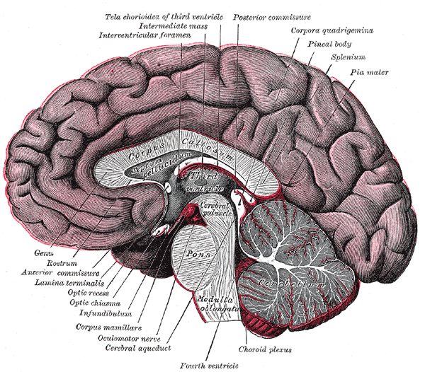
Mesal aspect of a brain sectioned in the median sagittal plane.
-

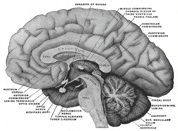
Median sagittal section of brain.
-
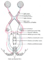
Scheme showing central connections of the optic nerves and optic tracts.
-
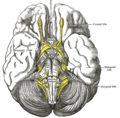
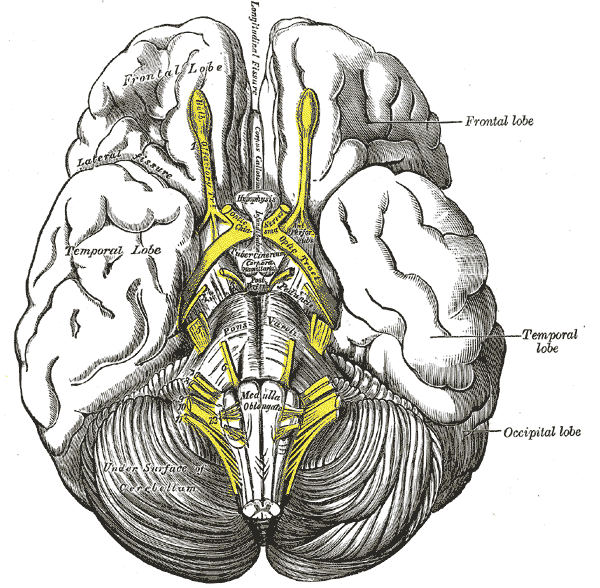
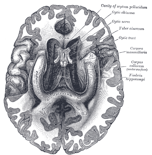
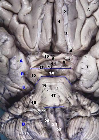
Base of brain.
-
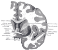
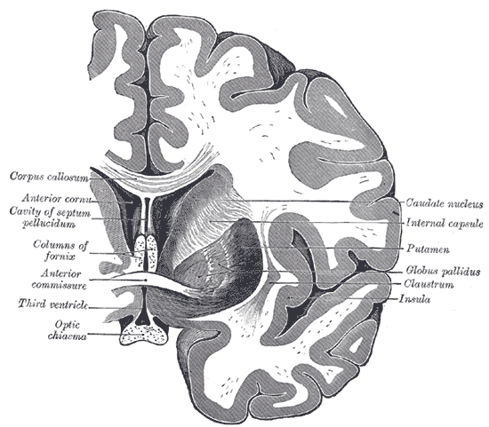
Coronal section of brain through anterior commissure.
-

The fornix and corpus callosum from below.
-
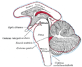
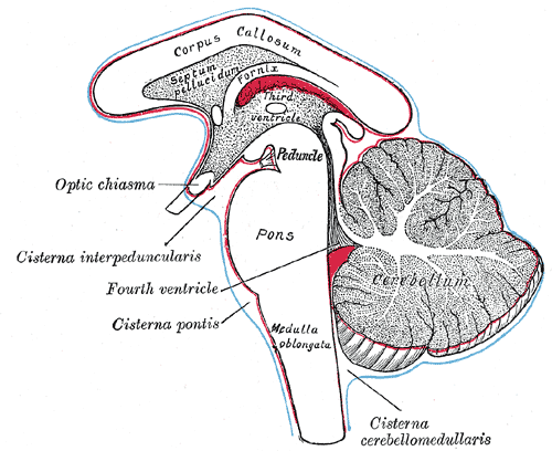
Diagram showing the positions of the three principal subarachnoid cisternæ.
-

The left optic nerve and the optic tracts.
-

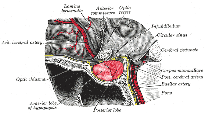
The hypophysis cerebri in position. Shown in sagittal section.
-

Median sagittal through the hypophysis of an adult monkey. Semidiagrammatic.
-

3D schematic representation of optic tracts
-
Human brainstem anterior view
See also
References
- ^ Colman, Andrew M. (2006). Oxford Dictionary of Psychology (2nd ed.). Oxford University Press. p. 530. ISBN 0198610351
- ^ "eye, human." Encyclopædia Britannica from Encyclopædia Britannica 2006 Ultimate Reference Suite DVD 2009
- ^ OMIA "Coat colour, albinism. Phenotype in cat (Felis catus).". OMIA. http://www.ncbi.nlm.nih.gov/entrez/query.fcgi?cmd=Retrieve&db=omia&dopt=Detailed&list_uids=418 OMIA. Retrieved 2009-09-26.
- ^ Guillery RW, Kaas JH (June 1973). "Genetic abnormality of the visual pathways in a "white" tiger". Science 180 (4092): 1287–9. doi:10.1126/science.180.4092.1287. PMID 4707916. http://www.sciencemag.org/cgi/pmidlookup?view=long&pmid=4707916.
- ^ Guillery RW (May 1974). "Visual pathways in albinos". Sci. Am. 230 (5): 44–54. doi:10.1038/scientificamerican0574-44. PMID 4822986.
- Jeffery G (October 2001). "Architecture of the optic chiasm and the mechanisms that sculpt its development". Physiol. Rev. 81 (4): 1393–414. PMID 11581492. http://physrev.physiology.org/cgi/content/full/81/4/1393.
External links
Nerves of head and neck: the cranial nerves and nuclei (TA A14.2.01, GA 9.855) olfactory (AON->I) optic (LGN->II) optic chiasm · optic tractoculomotor
(ON, EWN->III)trochlear (TN->IV) no significant branchestrigeminal
(PSN, TSN, MN, TMN->V)abducens (AN->VI) no significant branchesfacial (FMN, SN, SSN->VII) near origininside
facial canalvestibulocochlear
(VN, CN->VIII)glossopharyngeal
(NA, ISN, SN->IX)before jugular fossaafter jugular fossavagus
(NA, DNVN, SN->X)before jugular fossaafter jugular fossaaccessory (NA, SAN->XI) hypoglossal (HN->XII) Categories:- Cerebrum
- Visual system
- Neuroanatomy stubs
-


Wikimedia Foundation. 2010.