- Facial nerve
-
Nerve: Facial nerve 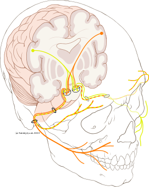
Cranial nerve VII 
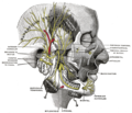
The nerves of the scalp, face, and side of neck. Latin nervus facialis Gray's subject #202 901 MeSH Facial+Nerve Cranial Nerves CN I – Olfactory CN II – Optic CN III – Oculomotor CN IV – Trochlear CN V – Trigeminal CN VI – Abducens CN VII – Facial CN VIII – Vestibulocochlear CN IX – Glossopharyngeal CN X – Vagus CN XI – Spinal Accessory CN XII – Hypoglossal The facial nerve is the seventh (VII) of twelve paired cranial nerves. It emerges from the brainstem between the pons and the medulla, and controls the muscles of facial expression, and functions in the conveyance of taste sensations from the anterior two-thirds of the tongue and oral cavity. It also supplies preganglionic parasympathetic fibers to several head and neck ganglia.
Contents
Course
The motor part of the facial nerve arises from the facial nerve nucleus in the pons while the sensory part of the facial nerve arises from the nervus intermedius.
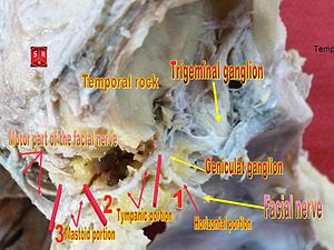
The motor part and sensory part of the facial nerve enters the petrous temporal bone into the internal auditory meatus (intimately close to the inner ear) then runs a tortuous course (including two tight turns) through the facial canal, emerges from the stylomastoid foramen and passes through the parotid gland, where it divides into five major branches. Though it passes through the parotid gland, it does not innervate the gland (This is the responsibility of cranial nerve IX, the glossopharyngeal nerve).
The facial nerve forms the geniculate ganglion prior to entering the facial canal.
Branches
Inside Skull
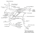
- Greater petrosal nerve - provides parasympathetic innervation to lacrimal gland, sphenoid sinus, frontal sinus, maxillary sinus, ethmoid sinus, nasal cavity, as well as special sensory taste fibers to the palate via the Vidian nerve.
- Nerve to stapedius - provides motor innervation for stapedius muscle in middle ear
- Chorda tympani - special sensory taste fibers for the anterior 2/3 of the tongue.
 Intrapetrous facial
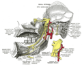
Outside skull (distal to stylomastoid foramen)
- Posterior auricular nerve - controls movements of some of the scalp muscles around the ear
- Branch to Posterior belly of Digastric and Stylohyoid muscle
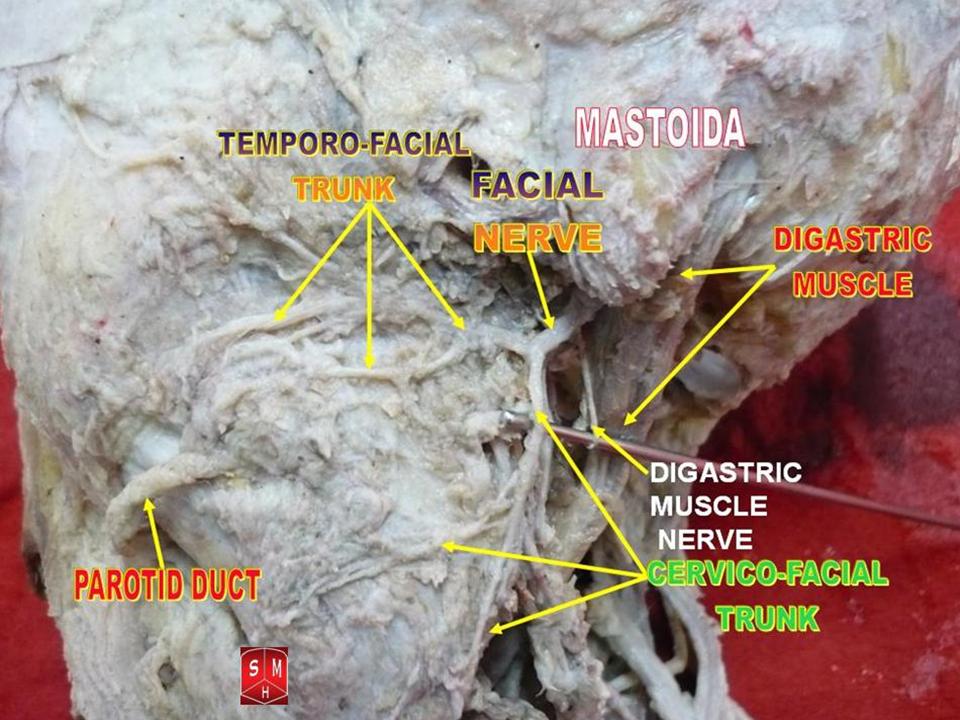
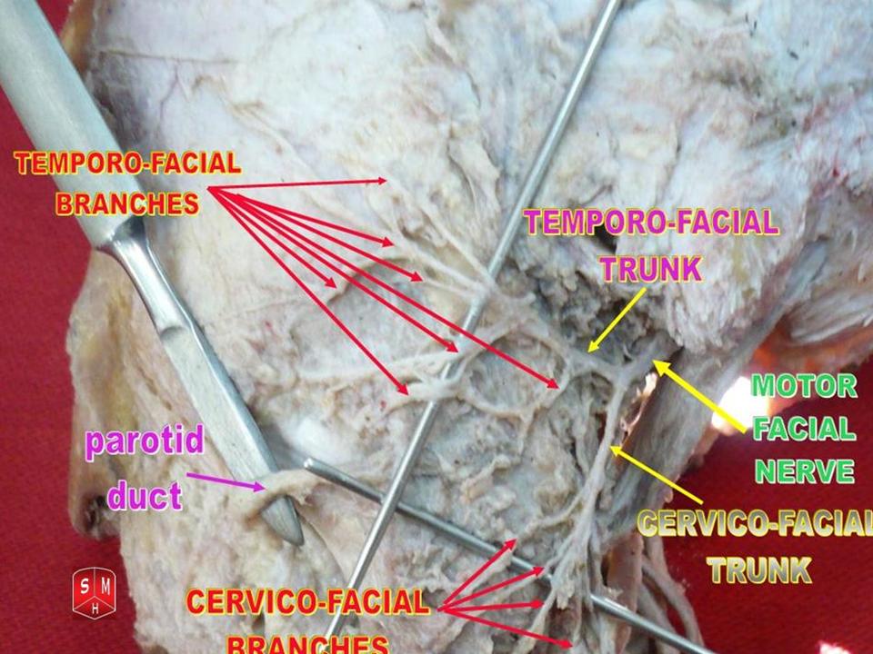
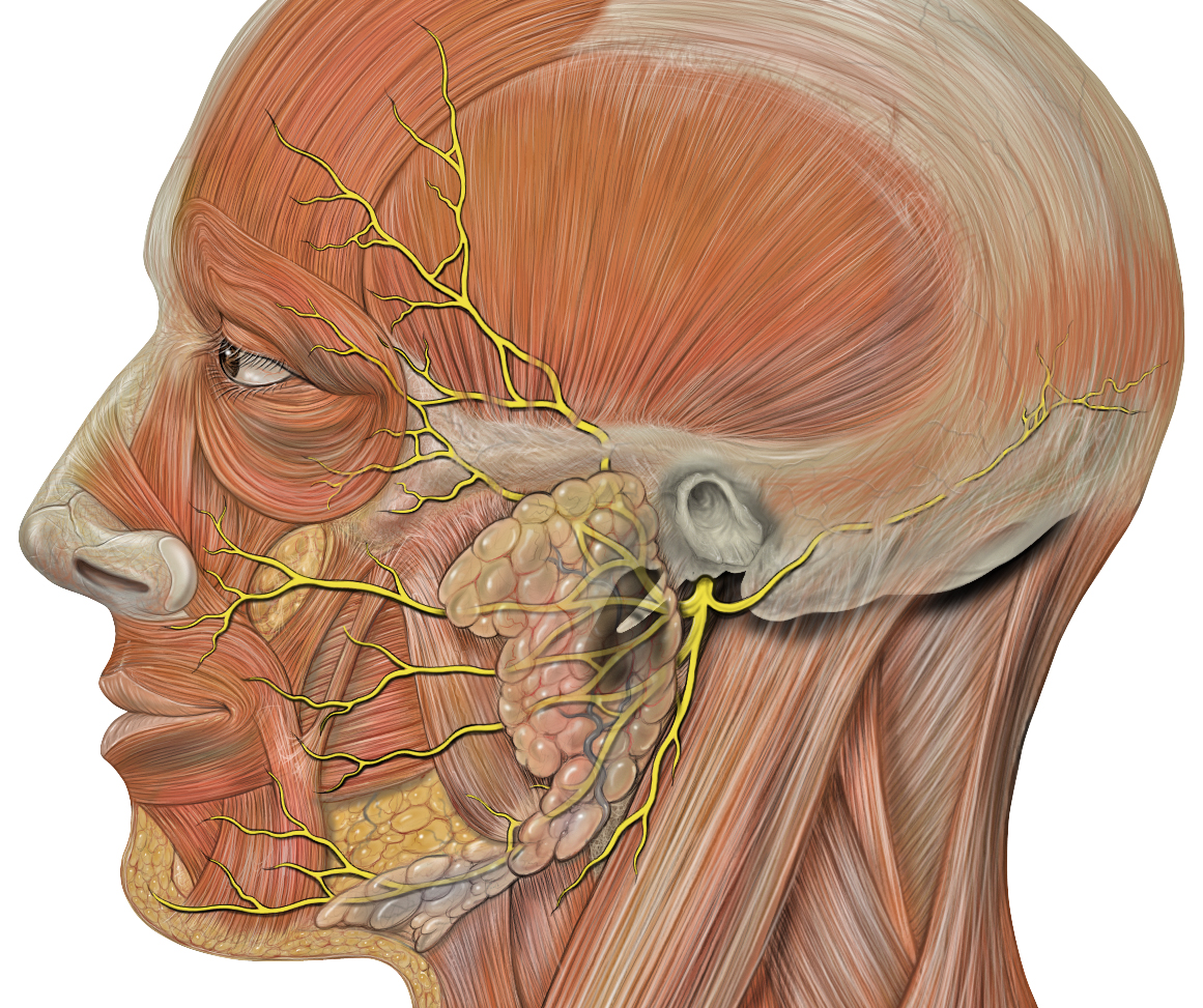
- Five major facial branches (in parotid gland) - from top to bottom:
- Temporal branch of the facial nerve
- Zygomatic branch of the facial nerve
- Buccal branch of the facial nerve
- Marginal mandibular branch of the facial nerve
- Cervical branch of the facial nerve
Intra operatively the facial nerve is recognised at 3 constant land marks:
1) At the tip of tragal cartilage where the nerve is 1cm deep and inferior
2) At the posterior belly of digastric by tracing this backwards to the tympanic plate the nerve can be found between these two structures
3) By locating the posterior facial vein at the inferior aspect of the gland where the marginal branch would be seen crossing it.A traditional mnemonic device for the five major branches of the facial nerve is:
"Ten Zebras Buggered My Cat"
"Today Zoe Bummed My Car"
"To Zanzibar By Motor Car"
"Ten Zimbabweans Burgled My Car"
"Tell Ziggy Bob Marley Called"
"Ten Zebras Bit My Cock"
"Two Zulus bombed my cat"
"Ten Zulus Bit My Chin"
"The Zoo Bought Monkey Clothes." and in Hindi "Tum Zyaada Buckbuck Mat K(C)aro".[citation needed]
A mnemonic device including the posterior auricular nerve is, "Perhaps A Tiny Zebra Bit My Cheek" or "Please Tell Ziggy Bob Marley Called".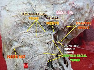 Facial nerve
Embryology
The facial nerve is developmentally derived from the hyoid arch (second pharyngeal branchial arch). The motor division of the facial nerve is derived from the basal plate of the embryonic pons, while the sensory division originates from the cranial neural crest.
Function
Efferent
Its main function is motor control of most of the muscles of facial expression. It also innervates the posterior belly of the digastric muscle, the stylohyoid muscle, and the stapedius muscle of the middle ear. All of these muscles are striated muscles of branchiomeric origin developing from the 2nd pharyngeal arch.
The facial also supplies parasympathetic fibers to the submandibular gland and sublingual glands via chorda tympani. Parasympathetic innervation serves to increase the flow of saliva from these glands. It also supplies parasympathetic innervation to the nasal mucosa and the lacrimal gland via the pterygopalatine ganglion.
The facial nerve also functions as the efferent limb of the corneal reflex.
Afferent
In addition, it receives taste sensations from the anterior two-thirds of the tongue via the chorda tympani, taste sensation is sent to the gustatory portion of the solitary nucleus. General sensation from the anterior two-thirds of tongue are supplied by afferent fibers of the third division of the fifth cranial nerve (V-3). These sensory (V-3) and taste (VII) fibers travel together as the lingual nerve briefly before the chorda tympani leaves the lingual Nerve to enter the middle temporal fossa via the petrotympanic fissure. It thus joins the rest of the facial nerve in the internal acoustic meatus before synapsing in the solitary nucleus. The cell bodies of the Chorda tympani reside in the geniculate ganglion, and these parasympathetic fibers synapse at the submandibular ganglion, attached To the lingual nerve.
The facial nerve also supplies a small amount of afferent innervation to the oropharynx below the palatine tonsil. There is also a small amount of cutaneous sensation carried by the nervus intermedius from the skin in and around the auricle (earlobe).
Location of Cell Bodies
The cell bodies for the facial nerve are grouped in anatomical areas called nuclei or ganglia. The cell bodies for the afferent nerves are found in the geniculate ganglion for taste sensation. The cell bodies for muscular efferent nerves are found in the facial motor nucleus whereas the cell bodies for the parasympathetic efferent nerves are found in the superior salivatory nucleus.
Infratemporal Paths
Upon reaching the temporal bone, the nerve's path can be divided into the internal auditory canal, labrynthine segment, intratympanic segment, and descending or vertical segment. The labrynthine segment is the narrowest portion of this pathway and is described to be approximately 0.7mm in diameter. The descending segment is the area where the branches of the chorda tympani and nerve to the stapedius branch from the facial nerve. The facial nerve eventually exits via the stylomastoid foramen to enter into the parotid where it branches into it's peripheral segments.
Pathology
People may suffer from acute facial nerve paralysis, which is usually manifested by facial paralysis. Bell's palsy is one type of idiopathic acute facial nerve paralysis, which is more accurately described as a multiple cranial nerve ganglionitis that involves the facial nerve, and most likely results from viral infection and also sometimes as a result of Lyme disease. Iatrogenic Bell's Palsy may also be as a result of an incorrectly placed dental local-anesthetic (Inferior alveolar nerve block). Although giving the appearance of a hemi-plegic stroke, effects dissipate with the drug.
Testing the facial nerve
Voluntary facial movements, such as wrinkling the brow, showing teeth, frowning, closing the eyes tightly (inability to do so is called lagophthalmos)[1] , pursing the lips and puffing out the cheeks, all test the facial nerve. There should be no noticeable asymmetry.
In an UMN lesion, called central seven, only the lower part of the face on the contralateral side will be affected, due to the bilateral control to the upper facial muscles (frontalis and orbicularis oculi).
Lower motor neuron lesions can result in a CNVII palsy (Bell's palsy is the term used to describe the idiopathic form of facial nerve palsy), manifested as both upper and lower facial weakness on the same side of the lesion.
Taste can be tested on the anterior 2/3 of the tongue. This can be tested with a swab dipped in a flavoured solution, or with electronic stimulation (similar to putting your tongue on a battery).
Corneal reflex. The afferent arc is mediated by the General Sensory afferents of the Trigeminal Nerve. The efferent arc occurs via the Facial Nerve. The reflex involves consensual blinking of both eyes in response to stimulation of one eye. This is due to the Facial Nerve's innervation of the muscles of facial expression, namely Orbicularis oculi, responsible for blinking. Thus, the corneal reflex effectively tests the proper functioning of both Cranial Nerves V and VII.
Additional images
-
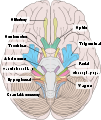
Inferior view of the human brain, with the cranial nerves labelled.
-
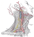
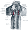
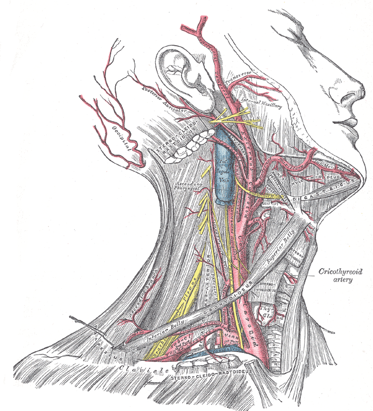
Superficial dissection of the right side of the neck, showing the carotid and subclavian arteries.
-
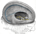
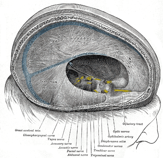
Dura mater and its processes exposed by removing part of the right half of the skull, and the brain.
-

Superficial dissection of brain-stem. Ventral view.
-
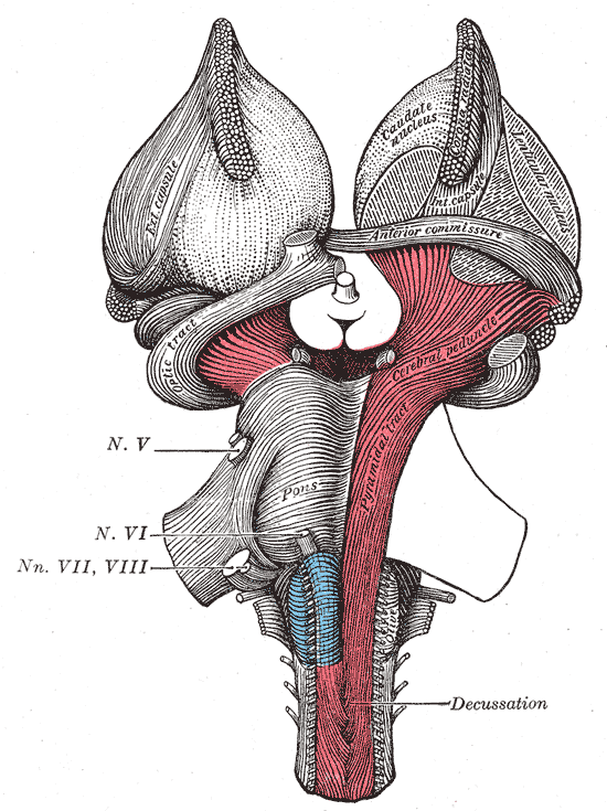
Hind- and mid-brains; postero-lateral view.
-
The sphenopalatine ganglion and its branches.
-
Mandibular division of the trifacial nerve.
-
Mandibular division of trifacial nerve, seen from the middle line.
-
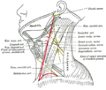
Plan of the facial and intermediate nerves and their communication with other nerves.
-
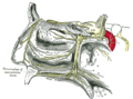
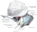
The course and connections of the facial nerve in the temporal bone.
-
Upper part of medulla spinalis and hind- and mid-brains; posterior aspect, exposed in situ.
-
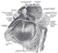
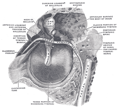
View of the inner wall of the tympanum (enlarged.)
-
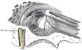
The right membrana tympani with the hammer and the chorda tympani, viewed from within, from behind, and from above.
-
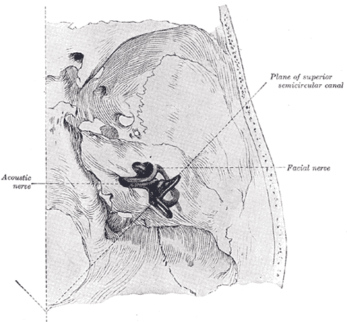
Position of the right bony labyrinth of the ear in the skull, viewed from above.
-

Facial nerve dissected
-
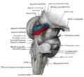
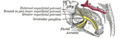
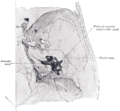
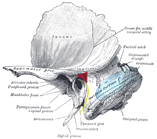
Left temporal bone showing surface markings for the tympanic antrum (red), transverse sinus (blue), and facial nerve (yellow).
-
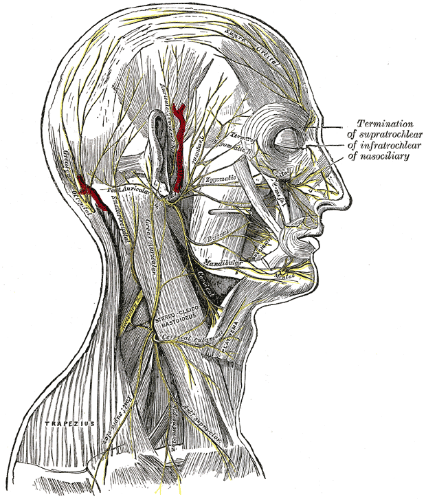
Side of neck, showing chief surface markings.
-

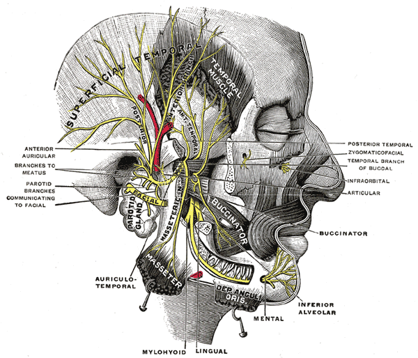
Head facial nerve branches
References
- ^ Kliniska Färdigheter: Informationsutbytet Mellan Patient Och Läkare, LINDGREN, STEFAN, ISBN 91-44-37271-X
External links
Nerves of head and neck: the cranial nerves and nuclei (TA A14.2.01, GA 9.855) olfactory (AON->I) optic (LGN->II) oculomotor
(ON, EWN->III)trochlear (TN->IV) no significant branchestrigeminal
(PSN, TSN, MN, TMN->V)abducens (AN->VI) no significant branchesfacial (FMN, SN, SSN->VII) near origininside
facial canalvestibulocochlear
(VN, CN->VIII)glossopharyngeal
(NA, ISN, SN->IX)before jugular fossaafter jugular fossavagus
(NA, DNVN, SN->X)before jugular fossaafter jugular fossaaccessory (NA, SAN->XI) hypoglossal (HN->XII) Categories:


Wikimedia Foundation. 2010.