- Medulla oblongata
-
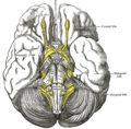
Brain: 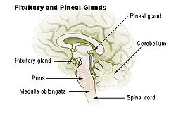
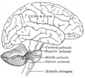
Medulla oblongata labeled at bottom left 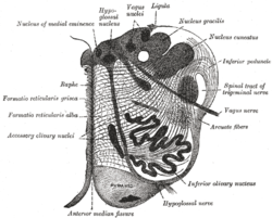
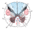
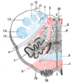
Section of the medulla oblongata at about the middle of the olive. Latin medulla oblongata Gray's subject #187 767 Part of Brain stem NeuroNames hier-695 MeSH Medulla+Oblongata NeuroLex ID birnlex_957 The medulla oblongata is the lower half of the brainstem. In discussions of neurology and similar contexts where no ambiguity will result, it is often referred to as simply the medulla. The medulla contains the cardiac, respiratory, vomiting and vasomotor centers and deals with autonomic, involuntary functions, such as breathing, heart rate and blood pressure.
Contents
Anatomy
Two parts: open and closed
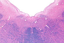 Micrograph of the posterior portion of the open part of the medulla oblongata, showing the fourth ventricle (top of image) and the nuclei of CN XII (medial) and CN X (lateral). H&E-LFB stain.
The medulla is often thought of as being in two parts:
- an open part or superior part where the dorsal surface of the medulla is formed by the fourth ventricle.
- a closed part or inferior part where the metacoel lies within the medulla oblongata.
Between the anterior median sulcus and the anterolateral sulcus
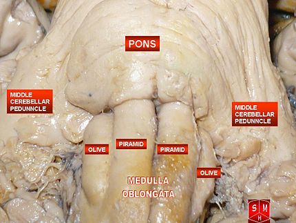
The region between the anterior median sulcus and the anterolateral sulcus is occupied by an elevation on either side known as the pyramid of medulla oblongata. This elevation is caused by the corticospinal tract.
In the lower part of the medulla some of these fibers cross each other thus obliterating the anterior median fissure. This is known as the decussation of the pyramids.
Some other fibers that originate from the anterior median fissure above the decussation of the pyramids and run laterally across the surface of the pons are known as the external arcuate fibers.
Between the anterolateral and posterolateral sulci
The region between the anterolateral and posterolateral sulcus in the upper part of the medulla is marked by a swelling known as the Olivary body.
It is caused by a large mass of gray matter known as the inferior olivary nucleus.
Between the posterior median sulcus and the posterolateral sulcus
The posterior part of the medulla between the posterior median sulcus and the posterolateral sulcus contains tracts that enter it from the posterior funiculus of the spinal cord. These are the fasciculus gracilis, lying medially next to the midline, and the fasciculus cuneatus, lying laterally.
These fasciculi end in rounded elevations known as the gracile and the cuneate tubercles. They are caused by masses of gray matter known as the nucleus gracilis and the nucleus cuneatus.
Just above the tubercles, the posterior aspect of the medulla is occupied by a triangular fossa, which forms the lower part of the floor of the fourth ventricle. The fossa is bounded on either side by the inferior cerebellar peduncle, which connects the medulla to the cerebellum.
Lower part
The lower part of the medulla, immediately lateral to the fasciculus cuneatus, is marked by another longitudinal elevation known as the tuberculum cinereum.
It is caused by an underlying collection of gray matter known as the spinal nucleus of the trigeminal nerve.
The gray matter of this nucleus is covered by a layer of nerve fibers that form the spinal tract of the trigeminal nerve.
Base
The base of the medulla is defined , the commissural fibers, crossing over from the ipsilateral side in the spinal cord to the contralateral side in the brain stem; below this is the spinal cord.
Functions
The medulla oblongata controls autonomic functions, and relays nerve signals between the brain and spinal cord. It is also responsible for controlling several major points and autonomic functions of the body:
- respiration – chemoreceptors
- cardiac center – sympathetic, parasympathetic system
- vasomotor center – baroreceptors
- reflex centers of vomiting, coughing, sneezing, and swallowing
Blood supply
Blood to the medulla is supplied by a number of arteries.
- Anterior spinal artery: The anterior spinal artery supplies the whole medial part of the medulla oblongata. A blockage (such as in a stroke) will injure the pyramidal tract, medial lemniscus, and the hypoglossal nucleus. This causes a syndrome called medial medullary syndrome.
- Posterior inferior cerebellar artery (PICA): The posterior inferior cerebellar artery, a major branch of the vertebral artery, supplies the posterolateral part of the medulla, where the main sensory tracts run and synapse. (As the name implies, it also supplies some of the cerebellum.)
- Direct branches of the vertebral artery: The vertebral artery supplies an area between the other two main arteries, including the nucleus solitarius and other sensory nuclei and fibers. Lateral medullary syndrome can be caused by occlusion of either the PICA or the vertebral arteries.
Embryology
The medulla oblongata forms from the lower (caudal) half of the embryonic rhombencephalon. Neuroblasts from the alar plate of the neural tube at this level will produce the sensory nuclei of the medulla. The basal plate neuroblasts will give rise to the motor nuclei.
- Alar plate neuroblasts give rise to:
- The solitary nucleus, which contains the general visceral afferent fibers (GVA) for taste, as well as the special visceral afferent (SVA) column.
- The spinal trigeminal nerve nuclei which contains the general somatic afferent column (GSA).
- The cochlear and vestibular nuclei, which contain the special somatic afferent (SSA) column.
- The inferior olivary nucleus, which relays to the cerebellum.
- The dorsal column nuclei, which contain the gracile and cuneate nuclei.
- Basal plate neuroblasts give rise to:
- The hypoglossal nucleus, which contains general somatic efferent fibers (GSE).
- The nucleus ambiguus, which form the special visceral efferent (SVE).
- The dorsal nucleus of vagus nerve and the inferior salivatory nucleus, both of which form the general visceral efferent fibers.
Evolution Theory
Both lampreys and hagfish possess a fully developed medulla oblongata.[1][2] Since these are both very similar to early agnathans, it has been suggested that the medulla evolved in these early fish, approximately 505 million years ago.[3]
Additional images
-
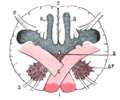
Section of the medulla\ oblongata through the lower part of the decussation of the pyramids
References
- ^ Nishizawa H et al. (1988). "Somatotopic organization of the primary sensory trigeminal neurons in the hagfish, Eptatretus burgeri.". J Comp Neurol. 267 (2): 281–95. doi:10.1002/cne.902670210. PMID 3343402. http://www.ncbi.nlm.nih.gov/pubmed/3343402.
- ^ Rovainen CM et al. (1985). "Respiratory bursts at the midline of the rostral medulla of the lamprey". J Comp Physiol A. 157 (3): 303–9.. doi:10.1007/BF00618120. PMID 3837091. http://www.ncbi.nlm.nih.gov/pubmed/3837091.
- ^ Haycock, Being and Perceiving
- Haycock DE (2011). Being and Perceiving. Manupod Press. ISBN 9780956962102. http://books.google.co.uk/books?id=fXFeQb1z6bsC.
External links
Nervous system (TA A14, GA 9) Central nervous system Brain: Rhombencephalon (Medulla oblongata, Pons, Cerebellum) • Mesencephalon • Prosencephalon (Diencephalon, Telencephalon)Peripheral nervous system Human brain: rhombencephalon, myelencephalon: medulla (TA A14.1.04, GA 9.767) Dorsal SurfacePosterior median sulcus · Posterolateral sulcus · Area postrema · Vagal trigone · Hypoglossal trigone · Medial eminenceafferent: GVA: VII,IX,X: Solitary/tract/Dorsal respiratory group · SVA: Gustatory nucleus · GSA: VIII-v (Lateral, Medial, Inferior)
efferent: GSE: XII · GVE: IX,X,XI: Ambiguus · SVE: X: Dorsal · IX: Inferior salivatory nucleusVentral Ventral respiratory group · Arcuate nucleus of medulla · Inferior olivary nucleus · Rostral ventromedial medullaSurfaceGrey: Raphe/
reticularCategories:
Wikimedia Foundation. 2010.

