- Rhombencephalon
-
Brain: Rhombencephalon 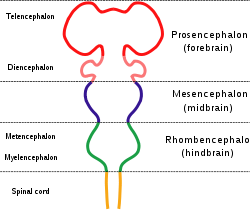
Diagram depicting the main subdivisions of the embryonic vertebrate brain. These regions will later differentiate into forebrain, midbrain and hindbrain structures. 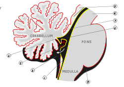
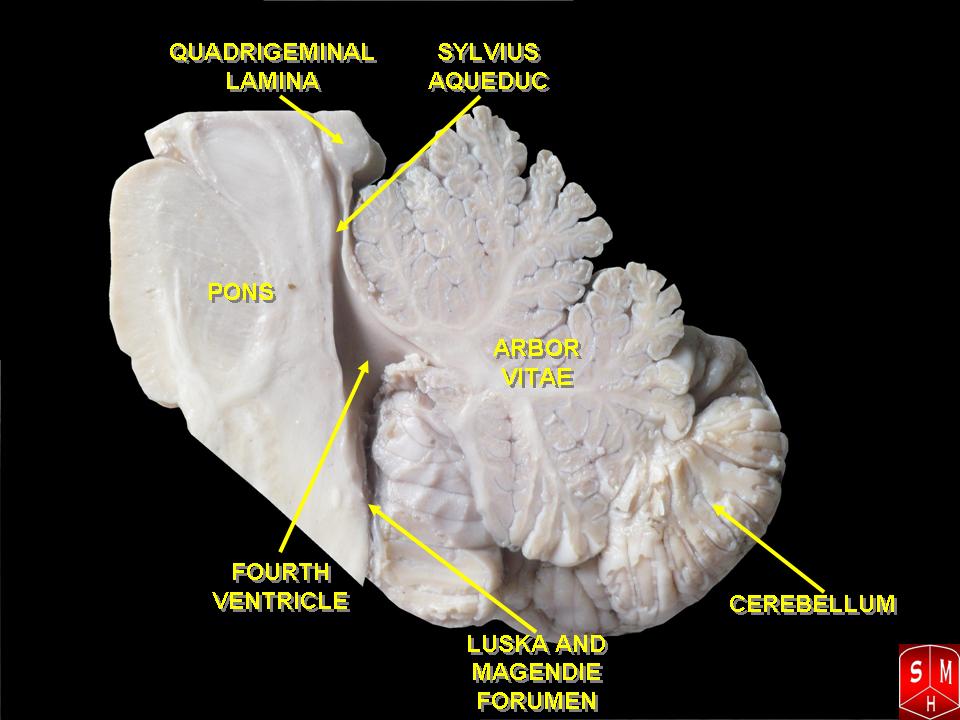
Scheme of roof of fourth ventricle. Gray's subject #187 767 NeuroNames hier-531 MeSH Rhombencephalon NeuroLex ID birnlex_942 The rhombencephalon (or hindbrain) is a developmental categorization of portions of the central nervous system in vertebrates.
 Rhombencephalon
The rhombencephalon can be subdivided in a variable number of transversal swellings called rhombomeres. In the human embryo eight rhombomeres can be distinguished, from caudal to rostral: Rh7-Rh1 and the isthmus (the most rostral rhombomere).
A rare disease of the rhombencephalon, "rhombencephalosynapsis," is characterized by a missing vermis resulting in a fused cerebellum. Patients generally present with cerebellar ataxia.
The caudal rhombencephalon has been generally considered as the initiation site for neural tube closure.[1]
Contents
Myelencephalon
Rhombomeres Rh7-Rh4 form the myelencephalon.
The myelencephalon forms the medulla oblongata in the adult brain; it contains:
- a portion of the fourth ventricle,
- the glossopharyngeal nerve (CN IX),
- vagus nerve (CN X),
- accessory nerve (CN XI),
- hypoglossal nerve (CN XII),
- and a portion of the vestibulocochlear nerve (CN VIII).
Metencephalon
Rhombomeres Rh3-Rh1 form the metencephalon.
The metencephalon is composed of the pons and the cerebellum; it contains:
- a portion of the fourth ventricle,
- the trigeminal nerve (CN V),
- abducens nerve (CN VI),
- facial nerve (CN VII),
- and a portion of the vestibulocochlear nerve (CN VIII).
Evolution
The hindbrain is homologous to a part of the arthropod brain known as the sub-oesophageal ganglion.[2] On this basis, it has been suggested that the hindbrain first evolved in the Urbilaterian - the last common ancestor of chordates and arthropods - between 570 and 555 million years ago.[2][3]
Additional images
-

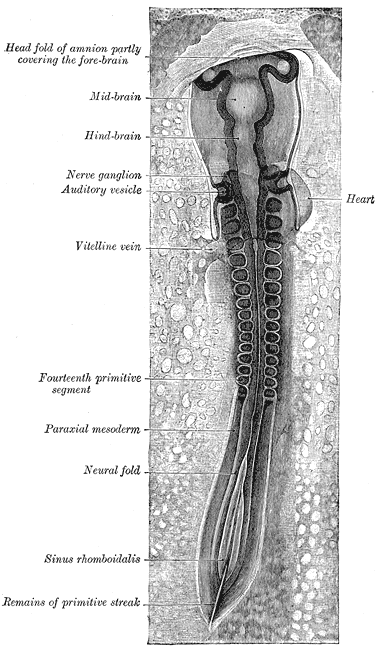
Chick embryo of thirty-three hours’ incubation, viewed from the dorsal aspect. X 30.
-
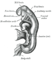
Embryo between eighteen and twenty-one days.
References
- Haycock DE (2011). Being and Perceiving. Manupod Press. ISBN 9780956962102. http://books.google.co.uk/books?id=fXFeQb1z6bsC.
- ^ SpringerLink - Journal Article
- ^ a b Ghysen A (2003). "The origin and evolution of the nervous system". Int. J. Dev. Biol. 47 (7–8): 555–62. PMID 14756331. http://www.ijdb.ehu.es/web/paper.php?doi=14756331.
- ^ Haycock, DE Being and Perceiving
External links
Nervous system (TA A14, GA 9) Central nervous system Brain: Rhombencephalon (Medulla oblongata, Pons, Cerebellum) • Mesencephalon • Prosencephalon (Diencephalon, Telencephalon)Peripheral nervous system Human brain: rhombencephalon, myelencephalon: medulla (TA A14.1.04, GA 9.767) Dorsal SurfacePosterior median sulcus · Posterolateral sulcus · Area postrema · Vagal trigone · Hypoglossal trigone · Medial eminenceafferent: GVA: VII,IX,X: Solitary/tract/Dorsal respiratory group · SVA: Gustatory nucleus · GSA: VIII-v (Lateral, Medial, Inferior)
efferent: GSE: XII · GVE: IX,X,XI: Ambiguus · SVE: X: Dorsal · IX: Inferior salivatory nucleusGrey: otherWhite: Sensory/ascendingWhite: Motor/descendingVentral White: Motor/descendingVentral respiratory group · Arcuate nucleus of medulla · Inferior olivary nucleus · Rostral ventromedial medullaSurfaceGrey: Raphe/
reticularHuman brain, rhombencephalon, metencephalon: pons (TA A14.1.05.101–604, GA 9.785) Dorsal/
(tegmentum)SurfaceWhite: Sensory/ascendingTrapezoid body/VIII · Trigeminal lemniscus (Dorsal trigeminal tract, Ventral trigeminal tract) · Medial lemniscus · Lateral lemniscus
MLF, III, IV and VI: Vestibulo-oculomotor fibers
Anterior trigeminothalamic tract · Central tegmental tractWhite: Motor/descendingICP (Vestibulocerebellar tract)
MLF, III, IV and VI: Vestibulospinal tract (Medial vestibulospinal tract, Lateral vestibulospinal tract)Other greyVentral/
(base)White: Motor/descendingSurfaceBasilar sulcusOther grey: Raphe/
reticularVentricular system, rhombencephalon, met- and myel-: fourth ventricle (TA A14.1.05.701–726, GA 9.797) Roof (dorsal) rostral: Superior medullary velum (Frenulum)
caudal: Inferior medullary velum · Taenia of fourth ventricleFloor/rhomboid fossa (ventral) rostral (pons): Facial colliculus · Locus coeruleus
caudal (medulla}: Vagal trigone · Hypoglossal trigone · Area postrema · Obex
Medial eminence · Sulcus limitansApertures Other Tela chorioidea of fourth ventricle · FastigiumHuman brain, rhombencephalon, metencephalon: cerebellum (TA 14.1.07, GA 9.788) Surface anatomy LobesMedial/lateralVermis: anterior (Central lobule, Culmen, Lingula) · posterior (Folium, Tuber, Uvula) · Vallecula of cerebellum
Hemisphere: anterior (Alar central lobule) · posterior (Biventer lobule, Cerebellar tonsil)Grey matter Molecular layer (Stellate cell, Basket cell)
Purkinje cell layer (Purkinje cell, Bergmann glia cell = Golgi epithelial cell)
Granule cell layer (Golgi cell, Granule cell, Unipolar brush cell)
Fibers: Mossy fibers · Climbing fiber · Parallel fiberWhite matter InternalPedunclesInferior (medulla): Dorsal spinocerebellar tract · Olivocerebellar tract · Cuneocerebellar tract · Juxtarestiform body (Vestibulocerebellar tract)
Middle (pons): Pontocerebellar fibers
Superior (midbrain): Ventral spinocerebellar tract · Dentatothalamic tract · Trigeminocerebellar fibersCategories:- Neuroanatomy
- Brainstem
- Central nervous system
- Cranial nerves
- Developmental biology
- Neuroanatomy stubs


Wikimedia Foundation. 2010.