- Dorsal cochlear nucleus
-
Brain: Dorsal cochlear nucleus 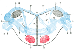
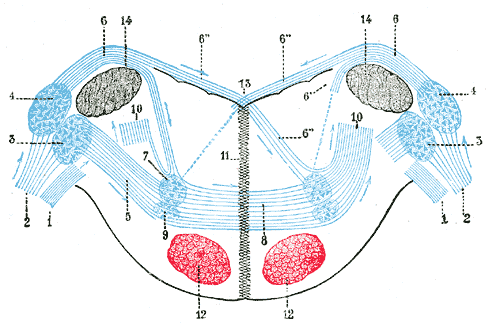
Dorsal cochlear nucleus is #4, at upper left 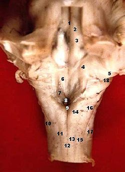
Human caudal brainstem posterior view description.JPG (Nucleus cochlearis dorsalis is #5) Latin nucleus cochlearis posterior NeuroNames hier-718 NeuroLex ID birnlex_2569 The dorsal cochlear nucleus (DCN, also known as the "tuberculum acousticum"), is a cortex-like structure on the dorso-lateral surface of the brainstem. Along with the ventral cochlear nucleus, it forms the cochlear nucleus, where all auditory nerve fibers from the cochlea form their first synapses.
Contents
Anatomy
The DCN differs from the ventral portion of the CN as it not only projects to the central nucleus of the Inferior Colliculus (ICC) but also receives efferent innervation from auditory cortex, superior olivary complex and inferior colliculus. The architecture and wiring of the DCN is similar to that of the cerebellum, a concept that currently is important in theories of DCN function. Thus, the DCN is thought to be involved with more complex auditory processing, rather than merely transferring information.
The fusiform (also called pyramidal) and giant cells are the principal cells of the DCN. There is no known physiological difference between these two cell types. These cells are the target of two different input systems. The first system arises from the auditory nerve, and carries acoustic information. The second set of inputs is relayed through a set of small cells called "granule" cells in the cochlear nucleus. The granule cells in turn are the target of a number of different inputs, including both those involved in auditory processing and, at least in lower mammals, somatosensory inputs associated with the head, the ear, and the jaw.
Projections from DCN principal cells form the dorsal acoustic stria, which ultimately terminate in the ICC. This projection overlaps with that of the LSO in a well-defined manner[1], where they form the primary excitatory input for ICC type O units[2]
Physiology
Principal cells in the DCN have very complex frequency intensity tuning curves. Classified as cochlear nucleus type IV cells[3], the firing rate may be very rapid in response to a low intensity sound at one frequency and then fall below the spontaneous rate with only a small increment in stimulus frequency or intensity. The firing rate may then increase with another increment in intensity or frequency. Type IV cells are excited by wide band noise, and particularly excited by a noise-notch stimulus directly below the cell's best frequency (BF).
While the VCN bushy cells aid in the location of a sound stimulus on the horizontal axis via their inputs to the superior olivary complex, type IV cells may participate in localization of the sound stimulus on the vertical axis. The pinna selectively amplifies frequencies, resulting in reduced sound energy at specific frequencies in certain regions of space. The complicated firing patterns of type IV cells makes them especially suited to detecting these notches, and with the combined power of these two localization systems, an ordinary person can locate where a firework explodes without the use of his eyes.
Somatosensory inputs inhibit type IV cell activity, possibly silencing their activity during head and pinna movements [4]. While this has not been studied extensively, it may play an important role in sound source localization in elevation. A similar effect is seen in the visual system in an effect known as change blindness.
Current auditory models of the DCN employ a two-inhibitor model. Type IV cells receive excitation directly from the auditory nerve, and are inhibited by type II (vertical) cells and a wide band inhibitor (onset-c cells).
References
- ^ Oliver, D. L., G. E. Beckius, et al. (1997). "Simultaneous anterograde labeling of axonal layers from lateral superior olive and dorsal cochlear nucleus in the inferior colliculus of cat." J Comp Neurol 382(2): 215-29.
- ^ Davis, K. A. (2002). "Evidence of a functionally segregated pathway from dorsal cochlear nucleus to inferior colliculus." J Neurophysiol 87(4): 1824-35.
- ^ Shofner, W. P. and E. D. Young (1985). "Excitatory/inhibitory response types in the cochlear nucleus: relationships to discharge patterns and responses to electrical stimulation of the auditory nerve." J Neurophysiol 54(4): 917-39.
- ^ Young, E. D., I. Nelken, et al. (1995). "Somatosensory effects on neurons in dorsal cochlear nucleus." J Neurophysiol 73(2): 743-65.
External links
Human brain, rhombencephalon, metencephalon: pons (TA A14.1.05.101–604, GA 9.785) Dorsal/
(tegmentum)SurfaceWhite: Sensory/ascendingTrapezoid body/VIII · Trigeminal lemniscus (Dorsal trigeminal tract, Ventral trigeminal tract) · Medial lemniscus · Lateral lemniscus
MLF, III, IV and VI: Vestibulo-oculomotor fibers
Anterior trigeminothalamic tract · Central tegmental tractWhite: Motor/descendingICP (Vestibulocerebellar tract)
MLF, III, IV and VI: Vestibulospinal tract (Medial vestibulospinal tract, Lateral vestibulospinal tract)Other greyVentral/
(base)White: Motor/descendingSurfaceBasilar sulcusOther grey: Raphe/
reticularAuditory and vestibular pathways Auditory inner ear: Hair cells → Spiral ganglion → Cochlear nerve VIII →
pons: Cochlear nuclei (Anterior, Dorsal) → Trapezoid body → Superior olivary nuclei →
midbrain: Lateral lemniscus → Inferior colliculi →
thalamus: Medial geniculate nuclei →
cerebrum: Acoustic radiation → Primary auditory cortexVestibular inner ear: Vestibular nerve VIII →
pons: Vestibular nuclei (Medial vestibular nucleus, Lateral vestibular nucleus)
cerebellum: Flocculonodular lobe
spinal cord: Vestibulospinal tract (Medial vestibulospinal tract, Lateral vestibulospinal tract)
thalamus: Ventral posterolateral nucleus
Vestibulo-oculomotor fibersM: EAR
anat(e/p)/phys/devp
noco/cong, epon
proc, drug(S2)
Categories:- Brainstem
- Neuroanatomy

Wikimedia Foundation. 2010.