- Cerebellopontine angle
-
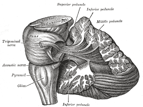
Brain: Cerebellopontine angle 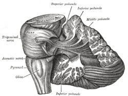
Dissection showing the projection fibers of the cerebellum 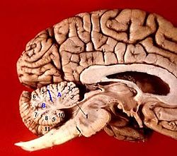
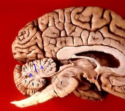
Human brain midsagittal view Latin Angulus pontocerebellaris The cerebellopontine angle is a structure at the margin of the cerebellum and pons.
Breast cancer can metastasize to the cerebellopontine angle.[1]
It can be a site for lipoma.[2]
It is where acoustic neuromas are usually found.
See also
References
- ^ Chiong Y, Mulroy L, Fleetwood IG, Younis T (October 2009). "Isolated metastasis to the cerebellopontine angle secondary to breast cancer". Can J Surg 52 (5): E213–4. PMC 2769124. PMID 19865565. http://www.cma.ca/multimedia/staticContent/HTML/N0/l2/cjs/vol-52/issue-5/pdf/pgE213.pdf.
- ^ Borges RS, Brito CC, Carvalho GA, Domingues RC, Gasparetto EL (June 2009). "Cerebellopontine angle lipomas: magnetic resonance imaging findings in two cases". Arq Neuropsiquiatr 67 (2B): 496–8. PMID 19623450. http://www.scielo.br/scielo.php?script=sci_arttext&pid=S0004-282X2009000300022&lng=en&nrm=iso&tlng=en.
External links
Human brain, rhombencephalon, metencephalon: pons (TA A14.1.05.101–604, GA 9.785) Dorsal/
(tegmentum)SurfaceCerebellopontine angle · Superior medullary velum · Sulcus limitans · Medial eminence · Facial colliculusWhite: Sensory/ascendingTrapezoid body/VIII · Trigeminal lemniscus (Dorsal trigeminal tract, Ventral trigeminal tract) · Medial lemniscus · Lateral lemniscus
MLF, III, IV and VI: Vestibulo-oculomotor fibers
Anterior trigeminothalamic tract · Central tegmental tractWhite: Motor/descendingICP (Vestibulocerebellar tract)
MLF, III, IV and VI: Vestibulospinal tract (Medial vestibulospinal tract, Lateral vestibulospinal tract)Other greyVentral/
(base)White: Motor/descendingSurfaceBasilar sulcusOther grey: Raphe/
reticularHuman brain, rhombencephalon, metencephalon: cerebellum (TA 14.1.07, GA 9.788) Surface anatomy LobesMedial/lateralVermis: anterior (Central lobule, Culmen, Lingula) · posterior (Folium, Tuber, Uvula) · Vallecula of cerebellum
Hemisphere: anterior (Alar central lobule) · posterior (Biventer lobule, Cerebellar tonsil)Grey matter Molecular layer (Stellate cell, Basket cell)
Purkinje cell layer (Purkinje cell, Bergmann glia cell = Golgi epithelial cell)
Granule cell layer (Golgi cell, Granule cell, Unipolar brush cell)
Fibers: Mossy fibers · Climbing fiber · Parallel fiberWhite matter InternalPedunclesInferior (medulla): Dorsal spinocerebellar tract · Olivocerebellar tract · Cuneocerebellar tract · Juxtarestiform body (Vestibulocerebellar tract)
Middle (pons): Pontocerebellar fibers
Superior (midbrain): Ventral spinocerebellar tract · Dentatothalamic tract · Trigeminocerebellar fibers
This anatomy article is a stub. You can help Wikipedia by expanding it.