- Cerebellar tonsil
-
Brain: Cerebellar tonsil 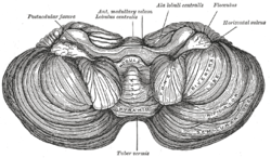
Anterior view of the cerebellum. (Tonsil visible at center right.) 
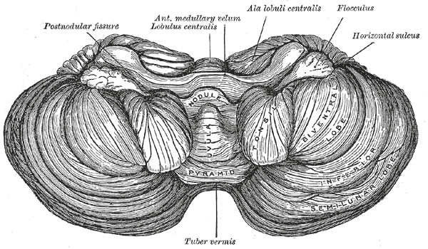
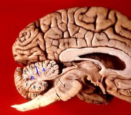
Sagittal section of the cerebellum, near the junction of the vermis with the hemisphere. (Tonsil visible at bottom center.) Latin tonsilla cerebelli Gray's subject #187 791 Part of Cerebellum Artery PICA NeuroNames hier-668 The cerebellar tonsil is analogous to a rounded lobule on the undersurface of each cerebellar hemisphere, continuous medially with the uvula of the Cerebellar vermis and superiorly by the flocculonodular lobe (source: Biology Online) Synonyms include: tonsilla cerebelli, amygdala cerebelli, the latter of which is not to be confused with the cerebral tonsils or amydgala nuclei located deep within the medial tempeoral lobes of the cerebral cortex. The flocculonodular lobe of the cerebellum which can also be confused for the cerebellar tonsils, is one of three lobes that make up the overall composition of the cerebellum. The cerebellum consists of three anatomical and functional lobes: anterior lobe, posterior lobe, and flocculonodular lobe.
Elongation of the cerebellar tonsils can, due to pressure, lead to this portion of the cerebellum to slip or be pushed through the foramen magnum of the skull resulting in a condition known as chiari malformation (CM).
Pathology
A Type I Arnold-Chiari malformation is a congenital anomaly of the brain in which the cerebellar tonsils are elongated and pushed down through the opening of the base of the skull (see foramen magnum), blocking the flow of cerebrospinal fluid (CSF). Also called tonsillar herniation. Although congenital, CM can also be induced due to physical head trauma.
Additional images
-

Human cerebellum anterior view
-
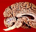
Human brain midsagittal view
This article was originally based on an entry from a public domain edition of Gray's Anatomy. As such, some of the information contained within it may be outdated.
Human brain, rhombencephalon, metencephalon: cerebellum (TA 14.1.07, GA 9.788) Surface anatomy LobesMedial/lateralVermis: anterior (Central lobule, Culmen, Lingula) · posterior (Folium, Tuber, Uvula) · Vallecula of cerebellum
Hemisphere: anterior (Alar central lobule) · posterior (Biventer lobule, Cerebellar tonsil)Grey matter Molecular layer (Stellate cell, Basket cell)
Purkinje cell layer (Purkinje cell, Bergmann glia cell = Golgi epithelial cell)
Granule cell layer (Golgi cell, Granule cell, Unipolar brush cell)
Fibers: Mossy fibers · Climbing fiber · Parallel fiberWhite matter InternalPedunclesInferior (medulla): Dorsal spinocerebellar tract · Olivocerebellar tract · Cuneocerebellar tract · Juxtarestiform body (Vestibulocerebellar tract)
Middle (pons): Pontocerebellar fibers
Superior (midbrain): Ventral spinocerebellar tract · Dentatothalamic tract · Trigeminocerebellar fibersCategories:- Cerebellum
- Neuroanatomy stubs
-

Wikimedia Foundation. 2010.