- Decussation of superior cerebellar peduncles
-
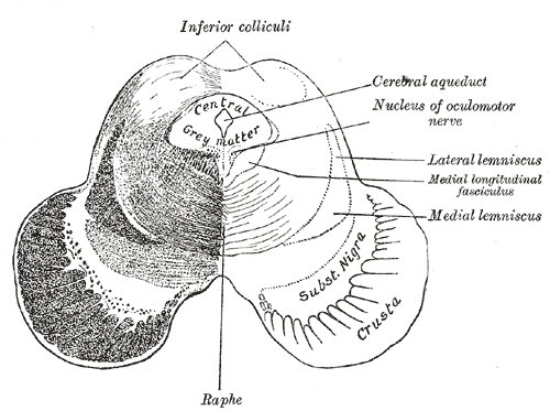
Brain: Decussation of superior cerebellar peduncles 
Transverse section of mid-brain at level of inferior colliculi. (Decussation not labeled, but region is visible.) Latin decussatio pedunculorum cerebellarium superiorum NeuroNames hier-518 NeuroLex ID birnlex_1089 The decussation of superior cerebellar peduncle is the portion of the superior cerebellar peduncle which crosses into the midbrain.
External links
- http://isc.temple.edu/neuroanatomy/lab/atlas/micn/
- http://www.neuroanatomy.wisc.edu/Bs97/TEXT/P18/sum.htm
- NIF Search - Decussation of superior cerebellar peduncle via the Neuroscience Information Framework
Human brain: mesencephalon (midbrain) (TA A14.1.06, GA 9.800) Tectum
(Dorsal)SurfaceWhite: Sensory/ascendingWhite: Motor/descendingPeduncle
(Ventral)White: Sensory/ascendinglemnisci (Medial, Lateral) · Ascending MLF (Vestibulo-oculomotor fibers) · Spinothalamic tract · Anterior trigeminothalamic tract · Dentatothalamic tractWhite: Motor/descendingGrey: otherPeriaqueductal gray/Raphe nuclei (Dorsal raphe nucleus)
Ventral tegmental area • Pedunculopontine nucleus • Red nucleus
riMLFBaseWhite: Motor/descendingSurfaceSuperior cerebellar peduncle (Decussation of superior cerebellar peduncles) · Interpeduncular fossaHuman brain, rhombencephalon, metencephalon: cerebellum (TA 14.1.07, GA 9.788) Surface anatomy LobesMedial/lateralVermis: anterior (Central lobule, Culmen, Lingula) · posterior (Folium, Tuber, Uvula) · Vallecula of cerebellum
Hemisphere: anterior (Alar central lobule) · posterior (Biventer lobule, Cerebellar tonsil)Grey matter Molecular layer (Stellate cell, Basket cell)
Purkinje cell layer (Purkinje cell, Bergmann glia cell = Golgi epithelial cell)
Granule cell layer (Golgi cell, Granule cell, Unipolar brush cell)
Fibers: Mossy fibers · Climbing fiber · Parallel fiberWhite matter InternalPedunclesInferior (medulla): Dorsal spinocerebellar tract · Olivocerebellar tract · Cuneocerebellar tract · Juxtarestiform body (Vestibulocerebellar tract)
Middle (pons): Pontocerebellar fibers
Superior (midbrain): Ventral spinocerebellar tract · Dentatothalamic tract · Trigeminocerebellar fibers
This anatomy article is a stub. You can help Wikipedia by expanding it.