- Cerebral peduncle
-
Brain: Cerebral peduncle 
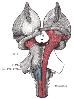
Superficial dissection of brain-stem. Ventral view. ("Cerebral peduncle" visible in red at center-right.) 
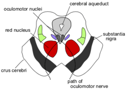
Section through superior colliculus showing path of oculomotor nerve. (Crus cerebri labeled at lower left.) Latin pedunculus cerebri Gray's subject #188 800 NeuroNames hier-478 MeSH Cerebral+Peduncle NeuroLex ID birnlex_1202 Mainly, the three common areas that give rise to the cerebral peduncles are the cortex, the spinal cord and the cerebellum.[1] The cerebral peduncle, by most classifications, is everything in the mesencephalon except the tectum.[citation needed] The region includes the midbrain tegmentum, crus cerebri and pretectum. By this definition, the cerebral peduncles are also known as the basis pedunculi, while the large ventral bundle of efferent fibers is referred to as the crus cerebri or the pes pedunculi. There are numerous nerve tracts located within this section of the brainstem. Of note, in the cerebral peduncular loop fibers from motor areas of the brain project to the cerebral peduncle and then project to various thalamic nuclei.
Important fibers running through the cerebral peduncles include the corticospinal tract and the corticobulbar tract, among others. In as much as the peduncles are an anatomic landmark, for details regarding the function of this area interested readers are referred to the individual referenced articles. On a broad scale, though, this area contains many nerve tracts conveying motor information to and from the brain to the rest of the body.
Contents
Additional images
See also
References
- ^ Saladin, Kenneth (2010), Anatomy & Physiology The Unity of Form and Function, New York, NY: McGraw-Hill Companies, Inc.
External links
Human brain: mesencephalon (midbrain) (TA A14.1.06, GA 9.800) Tectum
(Dorsal)SurfaceWhite: Sensory/ascendingWhite: Motor/descendingPeduncle
(Ventral)White: Sensory/ascendinglemnisci (Medial, Lateral) · Ascending MLF (Vestibulo-oculomotor fibers) · Spinothalamic tract · Anterior trigeminothalamic tract · Dentatothalamic tractWhite: Motor/descendingGrey: otherPeriaqueductal gray/Raphe nuclei (Dorsal raphe nucleus)
Ventral tegmental area • Pedunculopontine nucleus • Red nucleus
riMLFBaseWhite: Motor/descendingSurfaceCategories:
Wikimedia Foundation. 2010.


