- Cerebellar hemisphere
-
Brain: Cerebellar hemisphere 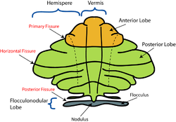
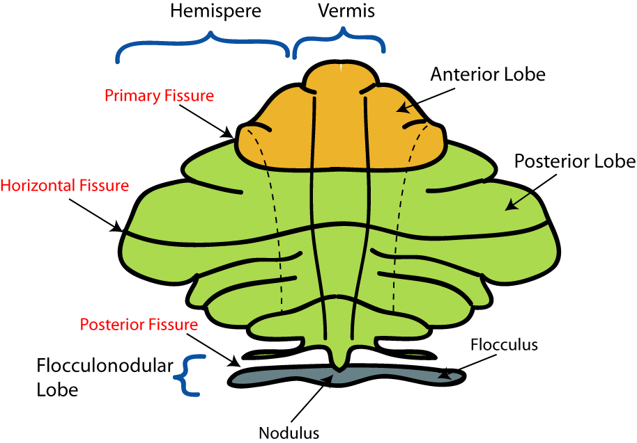
Schematic representation of the major anatomical subdivisions of the cerebellum. Superior view of an "unrolled" cerebellum, placing the vermis in one plane. 
Human cerebellum anterior view description (Cerebellar hemisphere is #8) Latin hemispherium cerebelli Gray's subject #187 788 NeuroNames ancil-214 NeuroLex ID birnlex_1575 The cerebellum consists of three parts, a median and two lateral, which are continuous with each other, and are substantially the same in structure. The median portion is constricted, and is called the vermis, from its annulated appearance which it owes to the transverse ridges and furrows upon it; the lateral expanded portions are named the hemispheres.
Portions
The "intermediate hemisphere" is also known as the "spinocerebellum".
The "lateral hemisphere" is also known as the "pontocerebellum".
The lateral hemisphere is considered the portion of the cerebellum to develop most recently.[1]
References
External links
- Atlas of anatomy at UMich n2a3p2
- NIF Search - Cerebellar Hemisphere via the Neuroscience Information Framework
This article was originally based on an entry from a public domain edition of Gray's Anatomy. As such, some of the information contained within it may be outdated.
Human brain, rhombencephalon, metencephalon: cerebellum (TA 14.1.07, GA 9.788) Surface anatomy LobesMedial/lateralVermis: anterior (Central lobule, Culmen, Lingula) · posterior (Folium, Tuber, Uvula) · Vallecula of cerebellum
Hemisphere: anterior (Alar central lobule) · posterior (Biventer lobule, Cerebellar tonsil)Grey matter Molecular layer (Stellate cell, Basket cell)
Purkinje cell layer (Purkinje cell, Bergmann glia cell = Golgi epithelial cell)
Granule cell layer (Golgi cell, Granule cell, Unipolar brush cell)
Fibers: Mossy fibers · Climbing fiber · Parallel fiberWhite matter InternalPedunclesInferior (medulla): Dorsal spinocerebellar tract · Olivocerebellar tract · Cuneocerebellar tract · Juxtarestiform body (Vestibulocerebellar tract)
Middle (pons): Pontocerebellar fibers
Superior (midbrain): Ventral spinocerebellar tract · Dentatothalamic tract · Trigeminocerebellar fibersCategories:- Neuroanatomy stubs
- Cerebellum
Wikimedia Foundation. 2010.