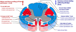
- Anterior corticospinal tract
-
Anterior corticospinal tract 
Anterior corticospinal tract seen in red at bottom center in figure (text tag found at upper left). 
Decussation of pyramids. Scheme showing passage of various fasciculi from medulla spinalis to medulla oblongata. a. Pons. b. Medulla oblongata. c. Decussation of the pyramids. d. Section of cervical part of medulla spinalis. 1. Anterior cerebrospinal fasciculus (in red). 2. Lateral cerebrospinal fasciculus (in red). 3. Sensory tract (fasciculi gracilis et cuneatus) (in blue). 3’. Gracile and cuneate nuclei. 4. Antero-lateral proper fasciculus (in dotted line). 5. Pyramid. 6. Lemniscus. 7. Medial longitudinal fasciculus. 8. Ventral spinocerebellar fasciculus (in blue). 9. Dorsal spinocerebellar fasciculus (in yellow). Latin tractus corticospinalis anterior, fasciculus cerebrospinalis anterior Gray's subject #185 759 The anterior corticospinal tract (also called the ventral corticospinal tract, medial corticospinal tract, direct pyramidal tract, or anterior cerebrospinal fasciculus) is a small bundle of descending fibers that connect the cerebral cortex to the spinal cord. It is usually small, varying inversely in size with the lateral corticospinal tract, which is the main part of the corticospinal tract.
It lies close to the anterior median fissure, and is present only in the upper part of the medulla spinalis; gradually diminishing in size as it descends, it ends about the middle of the thoracic region.
It consists of descending fibers which arise from cells in the motor area of the ipsilateral cerebral hemisphere, and which, as they run downward in the medulla spinalis, cross in succession through the anterior white commissure to the opposite side, where they end, either directly or indirectly, by arborizing around the motor neurons in the anterior column.
A few of its fibers pass to the lateral column of the same side and to the gray matter at the base of the posterior column.[citation needed]
They conduct voluntary motor impulses from the precentral gyrus to the motor centers of the cord.
Additional images
External links
- NeuroNames hier-799
- 745209857 at GPnotebook
- Anterior+corticospinal+tract at eMedicine Dictionary
- Overview at thebrain.mcgill.ca
This article was originally based on an entry from a public domain edition of Gray's Anatomy. As such, some of the information contained within it may be outdated.
Human brain: rhombencephalon, myelencephalon: medulla (TA A14.1.04, GA 9.767) Dorsal SurfacePosterior median sulcus · Posterolateral sulcus · Area postrema · Vagal trigone · Hypoglossal trigone · Medial eminenceafferent: GVA: VII,IX,X: Solitary/tract/Dorsal respiratory group · SVA: Gustatory nucleus · GSA: VIII-v (Lateral, Medial, Inferior)
efferent: GSE: XII · GVE: IX,X,XI: Ambiguus · SVE: X: Dorsal · IX: Inferior salivatory nucleusGrey: otherWhite: Sensory/ascendingWhite: Motor/descendingVentral White: Motor/descendingVentral respiratory group · Arcuate nucleus of medulla · Inferior olivary nucleus · Rostral ventromedial medullaSurfaceGrey: Raphe/
reticularAnatomy of torso (primarily): the spinal cord (TA 14.1.02, GA 9.749) External, dorsal Posterior median sulcus · Posterolateral sulcusGrey matter/
Rexed laminaeI–VI: Posterior hornI: Marginal nucleus · II: Substantia gelatinosa of Rolando · III+IV: Nucleus proprius · Spinal lamina V · Spinal lamina VIVII: Lateral hornVIII–IX: Anterior hornX: OtherWhite matter somatic/
ascending
(blue)Posterior/PCML: touch: Gracile · Cuneate
Lateral: proprioception: Spinocerebellar (Dorsal, Ventral) · pain/temp: Spinothalamic (Lateral, Anterior) · Posterolateral (Lissauer) · Spinotectal
Spinoreticular tract · Spino-olivary tractmotor/
descending
(red)Lateral: Corticospinal (Lateral) · Ep (Rubrospinal, Olivospinal)
Anterior: Corticospinal (Anterior) · Ep (Vestibulospinal, Reticulospinal, Tectospinal)bothExternal, ventral Anterior median fissure · Anterolateral sulcusExternal, general Categories:- Motor system
- Central nervous system pathways
- Neuroscience stubs
Wikimedia Foundation. 2010.


