- Marginal nucleus of spinal cord
-
Brain: Posteromarginal nucleus 
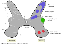
Medulla spinalis (Rexed lamina I labeled at upper left.) Latin nucleus marginalis medullae spinalis; lamina spinalis I NeuroNames ancil-984 The marginal nucleus of spinal cord, or posteromarginal nucleus, or Substantia Marginalis, Rexed lamina I, is located at the most dorsal aspect of the dorsal horn of the spinal cord. The neurons located here receive input primarily from Lissauer's tract and relay information related to pain and temperature sensation. Pain sensation relayed here cannot be modulated, e.g. pain from burning the skin.
Anatomy of torso (primarily): the spinal cord (TA 14.1.02, GA 9.749) External, dorsal Posterior median sulcus · Posterolateral sulcusGrey matter/
Rexed laminaeI–VI: Posterior hornI: Marginal nucleus · II: Substantia gelatinosa of Rolando · III+IV: Nucleus proprius · Spinal lamina V · Spinal lamina VIVII: Lateral hornVIII–IX: Anterior hornX: OtherWhite matter somatic/
ascending
(blue)Posterior/PCML: touch: Gracile · Cuneate
Lateral: proprioception: Spinocerebellar (Dorsal, Ventral) · pain/temp: Spinothalamic (Lateral, Anterior) · Posterolateral (Lissauer) · Spinotectal
Spinoreticular tract · Spino-olivary tractmotor/
descending
(red)Lateral: Corticospinal (Lateral) · Ep (Rubrospinal, Olivospinal)
Anterior: Corticospinal (Anterior) · Ep (Vestibulospinal, Reticulospinal, Tectospinal)bothExternal, ventral Anterior median fissure · Anterolateral sulcusExternal, general Categories:- Back anatomy
- Spinal cord
- Neuroscience stubs
Wikimedia Foundation. 2010.
