- Posterolateral tract
Infobox Anatomy
Name = PAGENAME
Latin = t. posterolateralis
GraySubject = 185
GrayPage = 762
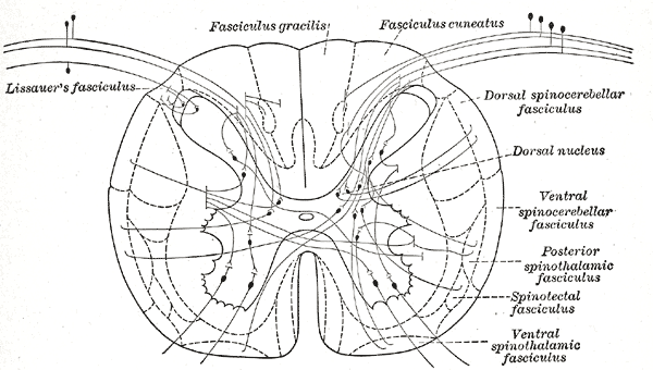
Caption = Diagram showing a few of the connections of afferent (sensory) fibers of the posterior root with the efferent fibers from the ventral column and with the various long ascending fasciculi. (Lissauer's fasciculus visible in upper left.)
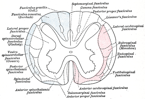
Caption2 = Diagram of the principal fasciculi of thespinal cord . (Lissauer's fasciculus visible in upper right.)
System =
Precursor =
MeshName =
MeshNumber =
DorlandsPre = t_15
DorlandsSuf = 12817084
The posterolateral tract (fasciculus of Lissauer, Lissauer's tract, tract of Lissauer, dorsolateral fasciculus, zone of Lissauer) is a small strand situated in relation to the tip of theposterior column close to the entrance of theposterior nerve roots .Composition and path
It contains centrally projecting axons carrying non-discriminative pain and temperature information (location, intensity and quality), which enter the spinal column ascend or descend one or two spinal segments in this tract before penetrating the grey mater of the dorsal horn where they synapse on second-order neurons. The axons of these second-order neurons cross the midline and ascend in the anterolateral quadrant of the contralateral half of the spinal cord, where they join the
spinothalamic tract . The second-order neurons ultimately synapse on neurons in the ventral posterior lateral nucleus (VPL) of thethalamus .It consists of fine fibers which do not receive their myelin sheaths until toward the close of
fetal life.In addition it contains great numbers of fine non-mylinated fibers derived mostly from the
dorsal roots but partlyendogenous in origin.These fibers are intimately related to the
substantia gelatinosa which is probably the terminal nucleus.The non-mylinated fibers ascend or descend for short distances not exceeding one or two segments, but most of them enter the substantia gelatinosa at or near the level of their origin.
Clinical significance
During a complete occlusion of the
ventral artery of the spinal cord , it is the only tract spared along with thedorsal column s.The posterolateral spinal tracts are involved withpernicious anemia .Eponym
The "tract of Lissauer" was named after German
neurologist Heinrich Lissauer (1861-1891).External links
*
* [http://instruct1.cit.cornell.edu/courses/psych396/student2006/the_biology_of_pain_mac_version/pathways.html Overview] atCornell University
Wikimedia Foundation. 2010.