Ventral spinocerebellar tract

Ventral spinocerebellar tract

Infobox Anatomy
Name = PAGENAME
Latin = tractus spinocerebellaris anterior, tractus spinocerebellaris ventralis
GraySubject = 185
GrayPage = 761


Caption = Ventral spinocerebellar tract is 4b, in blue at right.


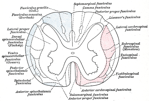
Caption2 = Diagram of the principal fasciculi of the spinal cord. (Ventral spinocerebellar fasciculus visible at center left.)
System =
Precursor =
MeshName =
MeshNumber =
DorlandsPre = t_15
DorlandsSuf = 12817184
The ventral spinocerebellar tract conveys proprioceptive information from the body to the cerebellum. It is part of the somatosensory system and runs in parallel with the dorsal spinocerebellar tract. Both these tracts involve two neurons and end up on the same side of the body.

The ventral tract (under L2/L3) gets its proprioceptive/fine touch/vibration information from a first order neuron, with its cell body in a dorsal ganglion. The axon runs via the fila radicularia to the dorsal horn of the grey matter. There it makes a synapse with the dendrites of two neurons: they send their axons bilateral to the ventral border of the lateral funiculi. The ventral spinocerebellar tract then enters the cerebellum via the superior cerebellar peduncle. This is in contrast with the dorsal spinocerebellar tract (C8 - L2/L3), which only has 1 unilateral axon that has its cell body in the Clarke's nuclei (only at the level of C8 - L2/L3). They enter the cerebellum via the inferior cerebellar peduncle.

Comparison with dorsal spinocerebellar tract

When the dorsal roots are cut in a cat performing a step cycle, the dorsal spinocerebellar tract has no activity with the loss of peripheral excitation, whereas the ventral spinocerebellar continues to show activity, implying that the dorsal spinocerebellar tract provides the spinocerebellum with sensory information during movement and the ventral spinocerebellar tract internally generated information about the movement. cite book |author=Jessell, Thomas M.; Kandel, Eric R.; Schwartz, James H. |title=Principles of neural science |publisher=McGraw-Hill |location=New York |year=2000 |pages= |isbn=0-8385-7701-6 |oclc= |doi=]

ynonyms

*anterior spinocerebellar tract
*Gowers' column
*Gowers' fasciculus
*Gowers' tract (named after Sir William Richard Gowers)


=Additional

References

External links

*
*


Wikimedia Foundation. 2010.

Игры ⚽ Нужен реферат?

Look at other dictionaries:

  • ventral spinocerebellar tract — n SPINOCEREBELLAR TRACT (b) …   Medical dictionary

  • spinocerebellar tract — n any of four nerve tracts which pass from the spinal cord to the cerebellum and of which two are situated on each side external to the crossed corticospinal tracts: a) a posterior tract on each side that arises from cells in the nucleus dorsalis …   Medical dictionary

  • Dorsal spinocerebellar tract — Posterior spinocerebellar tract is labeled in blue at right …   Wikipedia

  • Spinocerebellar tract — Infobox Brain Name = PAGENAME Latin = GraySubject = 185 GrayPage = 761 Caption = Spinocerebellar tract is #4, in blue at right. Caption2 = IsPartOf = Components = Artery = Vein = BrainInfoType = ancil BrainInfoNumber = 2137625602 MeshName =… …   Wikipedia

  • spinocerebellar tract ventral — tractus spinocerebellaris anterior …   Medical dictionary

  • tract — An elongated area, e.g., path, track, way. SEE ALSO: fascicle. SYN: tractus. [L. tractus, a drawing out] alimentary t. SYN: digestive t.. anterior corticospinal t. uncrossed fibers forming a small bundle in the anterior funiculus of the …   Medical dictionary

  • Cerebellothalamic tract — Also known as the dentatothalamic tract or the tractus cerebellothalamicus, is part of the superior cerebellar peduncle. It originates in the cerebellar nuclei, crosses completely in the decussation of the brachium conjunctivum, bypasses the red… …   Wikipedia

  • Dentatothalamic tract — Brain: Dentatothalamic tract Latin tractus dentatothalamicus NeuroNames hier 525 NeuroLex ID birnlex 1104 The dentatothalamic tract (or de …   Wikipedia

  • Olivocerebellar tract — Brain: Olivocerebellar tract Transverse section of medulla oblongata below the middle of the olive. (Cerebello olivary fibers visible at center right.) Latin t. olivocerebellaris NeuroNames …   Wikipedia

  • Corticospinal tract — Pyramidal tract redirects here. This page refers to the nerve fibres underlying the pyramids. For the actual area of the brain, Pyramids, see Pyramid of medulla oblongata. Brain: Corticospinal tract Deep dissection of brain stem. Lateral view. (… …   Wikipedia

Share the article and excerpts

Direct link
Do a right-click on the link above
and select “Copy Link”