- Motor cortex
-
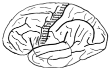
Motor cortex is a term that describes regions of the cerebral cortex involved in the planning, control, and execution of voluntary motor functions.
Contents
Anatomy of the motor cortex
The motor cortex can be divided into four main parts:
- the primary motor cortex (or M1), responsible for generating the neural impulses controlling execution of movement
- and the secondary motor cortices, including
- the posterior parietal cortex, responsible for transforming visual information into motor commands
- the premotor cortex, responsible for motor guidance of movement and control of proximal and trunk muscles of the body
- and the supplementary motor area (or SMA), responsible for planning and coordination of complex movements such as those requiring two hands.
Other brain regions outside the cortex are also of great importance to motor function, most notably the cerebellum and subcortical motor nuclei.
Early work on motor cortex function
In the 19th century Eduard Hitzig and Gustav Fritsch demonstrated that electrical stimulation of certain parts of the brain would result in muscular contraction on the opposite side of the body.[1]
In 1949 Canadian neurosurgeon Wilder Penfield developed a surgical procedure to relieve epilepsy. His initial procedure was to electrically probe the surface of the patient's cortex to find the problem area. During such investigations, he discovered that stimulation of Brodmann's area 4 readily elicited localised muscle twitches. Furthermore, there appeared to be a “motor map” of the body surface along the gyrus that comprises area 4. Area 4 is therefore now known as the primary motor cortex. Following this discovery, he discovered that stimulation of regions which are in front of the M1 caused more complicated movements; however, more electrical current was required to initiate movements from these areas. These 'premotor' cortical areas are located in Brodmann's area 6.
The motor cortical areas are now typically divided into three regions which have 2 different functional roles:
- primary motor cortex (M1)
- pre-motor area (PMA)
- supplementary motor area (SMA)
Penfield's experiments have made everything seem pretty straightforward: the purpose of M1 is to connect the brain to the lower motor neurons via the spinal cord in order to tell them which particular muscles need to contract. These upper motor neurons are found in layer 5 of the motor cortex and contain some of the largest cells in the brain (Betz cells whose cell bodies can be up to 100 micrometres in diameter. For comparison, rod photoreceptors are about 3 micrometres across). The descending axons of these layer 5 cells form the cortico-spinal or pyramidal tract. However, a single layer 5 forms synapses with many lower motor neurons which innervate different muscles. Furthermore, the same muscle is often represented over quite large regions of the brain's surface, and there is an overlap in the representation of different regions of the body.[citation needed] These facts mean that M1 neurons do not form simple connections with lower motor neurons. The activity of a single M1 neuron could cause contraction of more than one muscle; this suggests that M1 may not simply be coding the degree of contraction of individual muscles.
Non-activity responses in the motor cortex
Functional magnetic resonance imaging (fMRI) scans of persons reading words have shown that the act of reading a verb that refers to a face, arm, or leg action causes increased blood flow and activity in the motor cortex.[2] The areas of the motor cortex that are active correspond to sites of the motor cortex that are associated with that activity. For example, reading the word lick would increase blood flow in sites corresponding to tongue and mouth movements. While reading the verbs, blood flow also increases in premotor regions, Broca's area and Wernicke's area. Based on this information, it has been proposed that word understanding hinges on activation of interconnected brain areas that assimilate information about a particular word and its associated actions and sensations.[3]
References
- ^ Lennart Heimer (1995). The human brain and spinal cord: functional neuroanatomy and dissection guide. Springer-Verlag. ISBN 0-387-94227.
- ^ de Lafuente V, Romo R. (2004). "Language abilities of motor cortex". Neuron 41 (Jan. 22): 178–180. doi:10.1016/S0896-6273(04)00004-2. PMID 14741098.
- ^ Hauk O, Johnsrude I, Pulvermuller F (2004). "Somatotopic representation of action words in human motor and premotor cortex". Neuron 41 (Jan. 22): 301–307. doi:10.1016/S0896-6273(03)00838-9. PMID 14741110.
Canavero S. Textbook of therapeutic cortical stimulation. New York: Nova Science, 2009
Categories:
Wikimedia Foundation. 2010.

