- Poles of cerebral hemispheres
-
Brain: Poles of cerebral hemispheres 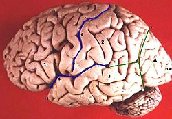
Human brain lateral view (9=Polus frontalis, 10=Polus occipitalis, 11=Polus temporalis) 
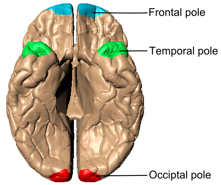
Lateral surface. (Frontal pole is approximately at 10, occipital pole is approximately at 17, and temporal pole is approximately at 38. Latin polus frontalis, polus occipitalis, polus temporalis Gray's subject #189 818 The anterior end of the hemisphere is named the frontal pole. (See also frontal lobe.)
The posterior end is named the occipital pole. (See also occipital lobe.)
The anterior end of the temporal lobe, the temporal pole. (See also temporal lobe.)
Gallery
-
 Poles of cerebral hemispheresFrontal poleTemporal poleOccipital pole
 -
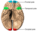
Cerebrum viewed from bottom.
See also
External links
- Photo at umanitoba.ca
- NeuroNames hier-123 (occipital pole)
- NeuroNames hier-38 (frontal pole)
- NeuroNames hier-108 (temporal pole)
This article was originally based on an entry from a public domain edition of Gray's Anatomy. As such, some of the information contained within it may be outdated.
Categories:- Neuroscience stubs
- Nervous system
-

Wikimedia Foundation. 2010.