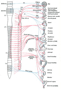
- Autonomic nervous system
-
Autonomic nervous system 
The autonomic nervous system
Blue = parasympathetic
Red = sympatheticLatin divisio autonomica systematis nervosi peripherici The autonomic nervous system (ANS or visceral nervous system) is the part of the peripheral nervous system that acts as a control system functioning largely below the level of consciousness, and controls visceral functions.[1] The ANS affects heart rate, digestion, respiration rate, salivation, perspiration, diameter of the pupils, micturition (urination), and sexual arousal. Whereas most of its actions are involuntary, some, such as breathing, work in tandem with the conscious mind.
It is classically divided into two subsystems: the parasympathetic nervous system (PSNS) and sympathetic nervous system (SNS).[1][2] Relatively recently, a third subsystem of neurons that have been named 'non-adrenergic and non-cholinergic' neurons (because they use nitric oxide as a neurotransmitter) have been described and found to be integral in autonomic function, particularly in the gut and the lungs.[3]
With regard to function, the ANS is usually divided into sensory (afferent) and motor (efferent) subsystems. Within these systems, however, there are inhibitory and excitatory synapses between neurons.
The enteric nervous system is sometimes considered part of the autonomic nervous system, and sometimes considered an independent system.
Contents
Anatomy
ANS innervation is divided into sympathetic nervous system and parasympathetic nervous system divisions. The sympathetic division has thoracolumbar “outflow”, meaning that the neurons begin at the thoracic and lumbar (T1-L2) portions of the spinal cord. The parasympathetic division has craniosacral “outflow”, meaning that the neurons begin at the cranial nerves (CN 3, CN7, CN 9, CN10) and sacral (S2-S4) spinal cord.
The ANS is unique in that it requires a sequential two-neuron efferent pathway; the preganglionic neuron must first synapse onto a postganglionic neuron before innervating the target organ. The preganglionic, or first, neuron will begin at the “outflow” and will synapse at the postganglionic, or second, neuron’s cell body. The post ganglionic neuron will then synapse at the target organ.
Sympathetic division
The sympathetic division (thoracolumbar outflow) consists of cell bodies in the lateral horn of spinal cord (intermediolateral cell columns) of the spinal cord from T1 to L2. These cell bodies are GVE (general visceral efferent) neurons and are the preganglionic neurons. There are several locations upon which preganglionic neurons can synapse for their postganglionic neurons:
- Paravertebral ganglia of the sympathetic chain (these run on either side of the vertebral bodies)
- Prevertebral ganglia (celiac ganglia, superior mesenteric ganglia, inferior mesenteric ganglia)
- Chromaffin cells of adrenal medulla (this is the one exception to the two-neuron pathway rule: synapse is direct onto cell bodies)
These ganglia provide the postganglionic neurons from which innervation of target organs follows. Examples of splanchnic (visceral) nerves are:
- Cervical cardiac nerves & thoracic visceral nerves which synapse in the sympathetic chain
- Thoracic splanchnic nerves (greater, lesser, least) which synapse in the prevertebral ganglion
- Lumbar splanchnic nerves which synapse in the prevertebral ganglion
- Sacral splanchnic nerves which synapse in the inferior hypogastric plexus
These all contain afferent (sensory) nerves as well, also known as GVA (general visceral afferent) neurons.
Parasympathetic division
The parasympathetic division (craniosacral outflow) consists of cell bodies from one of two locations: brainstem (Cranial Nerves III, VII, IX, X) or sacral spinal cord (S2, S3, S4). These are the preganglionic neurons, which synapse with postganglionic neurons in these locations:
- Parasympathetic ganglia of the head (Ciliary (CN III), Submandibular (CN VII), Pterygopalatine (CN VII), Otic (CN IX)
- In or near wall of organ innervated by Vagus (CN X), Sacral nerves (S2, S3, S4))
These ganglia provide the postganglionic neurons from which innervations of target organs follows. Examples are:
- The preganglionic parasympathetic splanchnic (visceral) nerves
- Vagus nerve, which wanders through the thorax and abdominal regions innervating, among other organs, the heart, lungs, liver and stomach
Sensory neurons
The sensory arm is made of “primary visceral sensory neurons” found in the peripheral nervous system (PNS), in “cranial sensory ganglia”: the geniculate, petrosal and nodose ganglia, appended respectively to cranial nerves VII, IX and X. These sensory neurons monitor the levels of carbon dioxide, oxygen and sugar in the blood, arterial pressure and the chemical composition of the stomach and gut content. (They also convey the sense of taste, a conscious perception). Blood oxygen and carbon dioxide are in fact directly sensed by the carotid body, a small collection of chemosensors at the bifurcation of the carotid artery, innervated by the petrosal (IXth) ganglion. Primary sensory neurons project (synapse) onto “second order” or relay visceral sensory neurons located in the medulla oblongata, forming the nucleus of the solitary tract (nTS), that integrates all visceral information. The nTS also receives input from a nearby chemosensory center, the area postrema, that detects toxins in the blood and the cerebrospinal fluid and is essential for chemically induced vomiting or conditional taste aversion (the memory that ensures that an animal which has been poisoned by a food never touches it again). All these visceral sensory informations constantly and unconsciously modulate the activity of the motor neurons of the ANS
Motor neurons
Motor neurons of the ANS are also located in ganglia of the PNS, called “autonomic ganglia”. They belong to three categories with different effects on their target organs (see below “Function”): sympathetic, parasympathetic and enteric.
Sympathetic ganglia are located in two sympathetic chains close to the spinal cord: the prevertebral and pre-aortic chains. Parasympathetic ganglia, in contrast, are located in close proximity to the target organ: the submandibular ganglion close to salivary glands, paracardiac ganglia close to the heart etc... Enteric ganglia, which as their name implies innervate the digestive tube, are located inside its walls and collectively contain as many neurons as the entire spinal cord, including local sensory neurons, motor neurons and interneurons. It is the only truly autonomous part of the ANS and the digestive tube can function surprisingly well even in isolation. For that reason the enteric nervous system has been called “the second brain”.
The activity of autonomic ganglionic neurons is modulated by “preganglionic neurons” (also called improperly but classically "visceral motoneurons") located in the central nervous system. Preganglionic sympathetic neurons are in the spinal cord, at thoraco-lumbar levels. Preganglionic parasympathetic neurons are in the medulla oblongata (forming visceral motor nuclei: the dorsal motor nucleus of the vagus nerve (dmnX), the nucleus ambiguus, and salivatory nuclei) and in the sacral spinal cord. Enteric neurons are also modulated by input from the CNS, from preganglionic neurons located, like parasympathetic ones, in the medulla oblongata (in the dmnX).
The feedback from the sensory to the motor arm of visceral reflex pathways is provided by direct or indirect connections between the nucleus of the solitary tract and visceral motoneurons.
Function
Sympathetic and parasympathetic divisions typically function in opposition to each other. But this opposition is better termed complementary in nature rather than antagonistic. For an analogy, one may think of the sympathetic division as the accelerator and the parasympathetic division as the brake. The sympathetic division typically functions in actions requiring quick responses. The parasympathetic division functions with actions that do not require immediate reaction. Consider sympathetic as "fight or flight" and parasympathetic as "rest and digest".
However, many instances of sympathetic and parasympathetic activity cannot be ascribed to "fight" or "rest" situations. For example, standing up from a reclining or sitting position would entail an unsustainable drop in blood pressure if not for a compensatory increase in the arterial sympathetic tonus. Another example is the constant, second to second modulation of heart rate by sympathetic and parasympathetic influences, as a function of the respiratory cycles. More generally, these two systems should be seen as permanently modulating vital functions, in usually antagonistic fashion, to achieve homeostasis. Some typical actions of the sympathetic and parasympathetic systems are listed below.
Sympathetic nervous system
Promotes a "fight or flight" response, corresponds with arousal and energy generation, and inhibits digestion.
- Diverts blood flow away from the gastro-intestinal (GI) tract and skin via vasoconstriction.
- Blood flow to skeletal muscles and the lungs is enhanced (by as much as 1200% in the case of skeletal muscles).
- Dilates bronchioles of the lung, which allows for greater alveolar oxygen exchange.
- Increases heart rate and the contractility of cardiac cells (myocytes), thereby providing a mechanism for the enhanced blood flow to skeletal muscles.
- Dilates pupils and relaxes the ciliary muscle to the lens, allowing more light to enter the eye and far vision.
- Provides vasodilation for the coronary vessels of the heart.
- Constricts all the intestinal sphincters and the urinary sphincter.
- Inhibits peristalsis.
- Stimulates orgasm.
Parasympathetic nervous system
Promotes a "rest and digest" response, promotes calming of the nerves return to regular function, and enhances digestion.
- Dilates blood vessels leading to the GI tract, increasing blood flow. This is important following the consumption of food, due to the greater metabolic demands placed on the body by the gut.
- The parasympathetic nervous system can also constrict the bronchiolar diameter when the need for oxygen has diminished.
- Dedicated cardiac branches of the Vagus and thoracic Spinal Accessory nerves impart Parasympathetic control of the Heart or Myocardium.
- During accommodation, the parasympathetic nervous system causes constriction of the pupil and contraction of the ciliary muscle to the lens, allowing for closer vision.
- The parasympathetic nervous system stimulates salivary gland secretion, and accelerates peristalsis, so, in keeping with the rest and digest functions, appropriate PNS activity mediates digestion of food and indirectly, the absorption of nutrients.
- Is also involved in erection of genitals, via the pelvic splanchnic nerves 2–4.
- Stimulates sexual arousal.
Neurotransmitters and pharmacology
At the effector organs, sympathetic ganglionic neurons release noradrenaline (norepinephrine), along with other cotransmitters such as ATP, to act on adrenergic receptors, with the exception of the sweat glands and the adrenal medulla:
- Acetylcholine is the preganglionic neurotransmitter for both divisions of the ANS, as well as the postganglionic neurotransmitter of parasympathetic neurons. Nerves that release acetylcholine are said to be cholinergic. In the parasympathetic system, ganglionic neurons use acetylcholine as a neurotransmitter, to stimulate muscarinic receptors.
- At the adrenal cortex, there is no postsynaptic neuron. Instead the presynaptic neuron releases acetylcholine to act on nicotinic receptors.
- Stimulation of the adrenal medulla releases adrenaline (epinephrine) into the bloodstream which will act on adrenoceptors, producing a widespread increase in sympathetic activity.
The following table reviews the actions of these neurotransmitters as a function of their receptors.
Circulatory system
Heart
Target Sympathetic (adrenergic) Parasympathetic (muscarinic) cardiac output β1, (β2): increases M2: decreases SA node: heart rate (chronotropic) β1, (β2) [4]: increases M2: decreases Atrial cardiac muscle: contractility (inotropic) β1, (β2)[4]: increases M2: decreases at AV node β1:
increases conduction
increases cardiac muscle automaticity [4]M2:
decreases conduction
Atrioventricular block [4]Ventricular cardiac muscle β1, (β2):
increases contractility (inotropic)
increases cardiac muscle automaticity [4]--- Blood vessels
Target Sympathetic (adrenergic) Parasympathetic (muscarinic) vascular smooth muscle in general α1:[5] contracts; β2:[5] relaxes M3: relaxes [4] renal artery α1[6]: constricts --- larger coronary arteries α1 and α2[7]: constricts [4] --- smaller coronary arteries β2:dilates [8] --- arteries to viscera α: constricts --- arteries to skin α: constricts --- arteries to brain α1[9]: constricts [4] --- arteries to erectile tissue α1[10]: constricts M3: dilates arteries to salivary glands α: constricts M3: dilates hepatic artery β2: dilates --- arteries to skeletal muscle β2: dilates --- Veins α1 and α2 [11] : constricts
β2: dilates--- Other
Target Sympathetic (adrenergic) Parasympathetic (muscarinic) platelets α2: aggregates --- mast cells - histamine β2: inhibits --- Respiratory system
Target Sympathetic (adrenergic) Parasympathetic (muscarinic) smooth muscles of bronchioles β2:[5] relaxes (major contribution)
α1: contracts (minor contribution)M3:[5] contracts The bronchioles have no sympathetic innervation, but are instead affected by circulating adrenaline [4]
Nervous system
Target Sympathetic (adrenergic) Parasympathetic (muscarinic) Pupil dilator muscle α1: Contracts
(causes mydriasis)- Iris sphincter muscle - M3: contracts
(causes miosis)Ciliary muscle β2: relaxes
(causes long-range focus)M3: contracts
(causes short-range focus)Digestive system
Target Sympathetic (adrenergic) Parasympathetic (muscarinic) salivary glands: secretions β: stimulates viscous, amylase secretions
α1: stimulates potassium secretionsM3: stimulates watery secretions lacrimal glands (tears) β: stimulates protein secretion [12] --- juxtaglomerular apparatus of kidney β1:[5] renin secretion --- parietal cells --- M1: Gastric acid secretion liver α1, β2: glycogenolysis, gluconeogenesis --- adipose cells β1,[5] β3: stimulates lipolysis --- GI tract (smooth muscle) motility α1, α2,[13] β2: decreases M3, (M1) [4]: increases sphincters of GI tract α1,[5] α2,[4] β2: contracts M3:[5] relaxes glands of GI tract no effect [4] M3: secretes Endocrine system
Target Sympathetic (adrenergic) Parasympathetic (muscarinic) pancreas (islets) α2: decreases insulin secretion from beta cells, increases glucagon secretion from alpha cells M3[14][15]: increases secretion of both insulin and glucagon.[14][15] adrenal medulla N (nicotinic ACh receptor): secretes epinephrine and norepinephrine --- Urinary system
Target Sympathetic (adrenergic) Parasympathetic (muscarinic) Detrusor urinae muscle of bladder wall β2:[5] relaxes M3:[5] contracts internal urethral sphincter α1:[5] contracts M3:[5] relaxes Reproductive system
Target Sympathetic (adrenergic) Parasympathetic (muscarinic) uterus α1: contracts (pregnant[4])
β2: relaxes (non-pregnant[4])--- genitalia α1: contracts (ejaculation) M3: erection Integumentary system
Target Sympathetic (muscarinic and adrenergic) Parasympathetic sweat gland secretions M:[5] stimulates (major contribution); α1: stimulates (minor contribution) --- arrector pili α1: stimulates --- References
- ^ a b "autonomic nervous system" at Dorland's Medical Dictionary
- ^ "eMedicine/Stedman Medical Dictionary Lookup!". http://www.emedicine.com/asp/dictionary.asp?keyword=autonomic+division+of+nervous+system. Retrieved 2008-11-30.[dead link]
- ^ "Nitric oxide is the endogenous neurotransmitter of bronchodilator nerves in humans Maria G. Belvisi, C. David Stretton, Magdi Yacouba, Peter J. Barnes". http://www.sciencedirect.com/science/article/pii/001429999290676U.
- ^ a b c d e f g h i j k l m n Rang, Dale, Ritter & Moore (2003). Pharmacology 5th ed.. Churchill Livingstone. p. 127. ISBN 0443071454.
- ^ a b c d e f g h i j k l m Page 37 in: Costanzo, Linda S. (2007). Physiology. Hagerstwon, MD: Lippincott Williams & Wilkins. ISBN 0-7817-7311-3.
- ^ Renal alpha-1 and alpha-2 adrenergic receptors: biochemical and pharmacological correlations JM Schmitz, RM Graham, A Sagalowsky and WA Pettinger
- ^ Coronary vasoconstriction mediated by alpha 1- and alpha 2-adrenoceptors in conscious dogs O. L. Woodman and S. F. Vatner
- ^ Rang, H. P. (2003). Pharmacology. Edinburgh: Churchill Livingstone. ISBN 0-443-07145-4. Page 270
- ^ Circulation & Lung Physiology I M.A.S.T.E.R. Learning Program, UC Davis School of Medicine
- ^ 1A-Adrenoceptors mediate contractions to phenylephrine in rabbit penile arteries J S Morton1, C J Daly1, V M Jackson2 and J C McGrath1: "1A-ARs could be utilized to aid the erectile response in male erectile dysfunction sufferers by reducing vasoconstriction of the penile arteries"
- ^ Elliott, J (1997). "Alpha-adrenoceptors in equine digital veins: Evidence for the presence of both alpha1 and alpha2-receptors mediating vasoconstriction". Journal of Veterinary Pharmacology & Therapeutics (Blackwell Publishings) 20 (4): 308–317. doi:10.1046/j.1365-2885.1997.00078.x. PMID 9280371. http://www.ingentaconnect.com/content/bsc/jvpt/1997/00000020/00000004/art00009;jsessionid=8lm5thggpj1x.alice?format=print.
- ^ Protein secretion induced by isoproterenol or pentoxifylline in lacrimal gland P. Mauduit, G. Herman and B. Rossignol
- ^ Sagrada, A; Fargeas, MJ; Bueno, L., A; Fargeas, MJ; Bueno, L (1987). "Involvement of alpha-1 and alpha-2 adrenoceptors in the postlaparotomy intestinal motor disturbances in the rat". Gut 28 (8): 955–959. doi:10.1136/gut.28.8.955. PMC 1433140. PMID 2889649. http://www.pubmedcentral.nih.gov/articlerender.fcgi?tool=pmcentrez&artid=1433140.
- ^ a b Page 47 in: Poretsky, Leonid (2010). Principles of diabetes mellitu. New York: Springer. ISBN 9780387098401.
- ^ a b Duttaroy, A.; Zimliki, C. L.; Gautam, D.; Cui, Y.; Mears, D.; Wess, J. (2004). "Muscarinic Stimulation of Pancreatic Insulin and Glucagon Release is Abolished in M3 Muscarinic Acetylcholine Receptor-Deficient Mice". Diabetes 53 (7): 1714–1720. doi:10.2337/diabetes.53.7.1714. PMID 15220195.
External links
- Receptors of the heart: Mnemonic
- Overview at arizona.edu
- ANS Medical Notes on rahulgladwin.com
- Explanations on the ANS by Dyansys, Inc.
Nervous system (TA A14, GA 9) Central nervous system Peripheral nervous system AutonomicCategories:
Wikimedia Foundation. 2010.
