- Spinal nerve
-
Nerve: Spinal nerve 
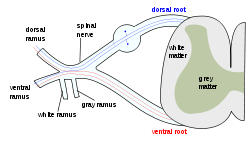
The formation of the spinal nerve from the dorsal and ventral roots 
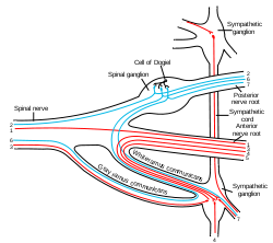
Scheme showing structure of a typical spinal nerve.
1. Somatic efferent.
2. Somatic afferent.
3,4,5. Sympathetic efferent.
6,7. Sympathetic afferent.Latin nervi spinales Gray's subject #208 916 Code TA A14.2.00.027 MeSH Spinal+nerves The term spinal nerve generally refers to a mixed spinal nerve, which carries motor, sensory, and autonomic signals between the spinal cord and the body. Humans have 31 left-right pairs of spinal nerves, each roughly corresponding to a segment of the vertebral column: 8 cervical spinal nerve pairs (C1-C8), 12 thoracic pairs (T1-T12), 5 lumbar pairs (L1-L5), 5 sacral pairs (S1-S5), and 1 coccygeal pair. The spinal nerves are part of the peripheral nervous system (PNS).
Contents
Anatomy
Each spinal nerve is formed by the combination of nerve fibers from the dorsal and ventral roots of the spinal cord. The dorsal roots carry afferent sensory axons, while the ventral roots carry efferent motor axons. The spinal nerve emerges from the spinal column through an opening (intervertebral foramen) between adjacent vertebrae. This is true for all spinal nerves except for the first spinal nerve pair, which emerges between the occipital bone and the atlas (the first vertebra).
Outside the vertebral column, the nerve divides into branches. The dorsal ramus contains nerves that serve the dorsal portions of the trunk carrying visceral motor, somatic motor, and sensory information to and from the skin and muscles of the back. The ventral ramus contains nerves that serve the remaining ventral parts of the trunk and the upper and lower limbs carrying visceral motor, somatic motor, and sensory information to and from the ventrolateral body surface, structures in the body wall, and the limbs. The meningeal branches (recurrent meningeal or sinuvertebral nerves) branch from the spinal nerve and re-enter the intervertebral foramen to serve the ligaments, dura, blood vessels, intervertebral discs, facet joints, and periosteum of the vertebrae. The rami communicantes contain autonomic nerves that serve visceral functions carrying visceral motor and sensory information to and from the visceral organs.
Some ventral rami merge with adjacent ventral rami to form a nerve plexus, a network of interconnecting nerves. Nerves emerging from a plexus contain fibers from various spinal nerves, which are now carried together to some target location. Major plexuses include the cervical, brachial, lumbar, and sacral plexuses.
Clinical significance
The muscles that one particular spinal root supplies are that nerve's myotome, and the dermatomes are the areas of sensory innervation on the skin for each spinal nerve. Lesions of one or more nerve roots result in typical patterns of neurologic defects (muscle weakness, abnormal sensation, changes in reflexes) that allow localization of the causeating lesion.
External links
- spinal+nerves at eMedicine Dictionary
References
- Blumenfeld H. 'Neuroanatomy Through Clinical Cases'. Sunderland, Mass: Sinauer Associates; 2002.
- Drake RL, Vogl W, Mitchell AWM. 'Gray's Anatomy for Students'. New York: Elsevier; 2005:69-70.
- Ropper AH, Samuels MA. 'Adams and Victor's Principles of Neurology'. Ninth Edition. New York: McGraw Hill; 2009.
Nervous system (TA A14, GA 9) Central nervous system Peripheral nervous system Nerves: spinal nerves (TA A14.2, GA 9.916) Cervical (8) Thoracic (12) T1, T2, T3, T4, T5, T6, T7, T8, T9, T10, T11, T12
anterior (Intercostal, Intercostobrachial – T2, Thoraco-abdominal nerves – T7–T11, Subcostal – T12) – posterior (Posterior branches of thoracic nerves)Lumbar (5)
anterior (Lumbar plexus, Lumbosacral trunk) · posterior (Posterior branches of the lumbar nerves, Superior cluneal L1–L3)Sacral (5) Coccygeal (1) Categories:- Peripheral nervous system
- Spinal nerves
Wikimedia Foundation. 2010.
