- Posterior ramus of spinal nerve
-
Nerve: Dorsal ramus of spinal nerve 
Diagram of the course and branches of a typical intercostal nerve. (Posterior division labeled at upper right.) 
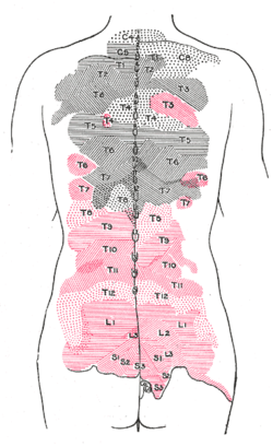
Areas of distribution of the cutaneous branches of the posterior divisions of the spinal nerves. The areas of the medial branches are in black, those of the lateral in red. Latin ramus posterior nervi spinalis Gray's subject #209 921 The posterior (or dorsal) branches (or divisions) of the spinal nerves are as a rule smaller than the anterior divisions. They are also referred to as the dorsal rami.
They are directed backward, and, with the exceptions of those of the first cervical, the fourth and fifth sacral, and the coccygeal, divide into medial and lateral branches for the supply of the muscles and skin of the posterior part of the trunk.
Shortly after a spinal nerve exits the intervertebral foramen, it branches into the dorsal ramus, ventral ramus, and rami communicantes. Each of these latter three structures carries both sensory and motor information. Because each spinal nerve carries both sensory and motor information, spinal nerves are referred to as “mixed nerves.” Dorsal rami carry visceral motor, somatic motor, and sensory information to and from the skin and deep muscles of the back.
Dorsal rami remain distinct from each other, and each innervates a narrow strip of skin and muscle along the back, more or less at the level from which the ramus leaves the spinal nerve.
The smaller, posteriorly-directed major terminal branch (with the ventral primary ramus) of all 31 pairs of mixed spinal nerves, formed at the intervertebral foramen and turning abruptly posteriorly to divide into lateral and medial branches, both of which will supply the deep (true) muscles of the back. The medial branch (rami medialis ) of the dorsal primary ramus also supplies articular branches to the zygopophyseal joints and the periosteum of the vertebral arch. In the neck and upper back, the medial branch continues through the deep and superficial back muscles to supply overlying skin; in the lower back, the lateral branch does this. Nomina Anatomica lists dorsal primary rami as "rami dorsales" for each group of spinal nerves: 1) cervical (nervorum cervicalium ), 2) thoracic (nervorum thoracicorum ), 3) lumbar (nervorum lumbalium ), 4) sacral (nervorum sacralium ), and 5) coccygeal (nervi coccygei ).[1]
Synonyms: ramus dorsalis nervorum spinalium, ramus dorsalis, rami posteriores nervorum spinalium, dorsal branch, posterior primary division.
Contents
References
See also
Additional images
External links
- posterior+ramus+of+spinal+nerve at eMedicine Dictionary
- terminologyanatplanes at The Anatomy Lesson by Wesley Norman (Georgetown University) (typicalspinalnerve)
Histology: nervous tissue (TA A14, GA 9.849, TH H2.00.06, H3.11) CNS GeneralGrey matter · White matter (Projection fibers · Association fiber · Commissural fiber · Lemniscus · Funiculus · Fasciculus · Decussation · Commissure) · meningesOtherPNS GeneralPosterior (Root, Ganglion, Ramus) · Anterior (Root, Ramus) · rami communicantes (Gray, White) · Autonomic ganglion (Preganglionic nerve fibers · Postganglionic nerve fibers)Myelination: Schwann cell (Neurolemma, Myelin incisure, Myelin sheath gap, Internodal segment)
Satellite glial cellNeurons/
nerve fibersPartsPerikaryon (Axon hillock)
Axon (Axon terminals, Axoplasm, Axolemma, Neurofibril/neurofilament)
Dendrite (Nissl body, Dendritic spine, Apical dendrite/Basal dendrite)TypesGSA · GVA · SSA · SVA
fibers (Ia, Ib or Golgi, II or Aβ, III or Aδ or fast pain, IV or C or slow pain)GSE · GVE · SVE
Upper motor neuron · Lower motor neuron (α motorneuron, γ motorneuron, β motorneuron)Termination SynapseCategories:- Spinal nerves
- Neuroscience stubs
Wikimedia Foundation. 2010.

