- Nissl body
-
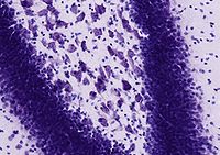 Image of a Nissl-stained histological section through the rodent hippocampus showing various classes of neurons.
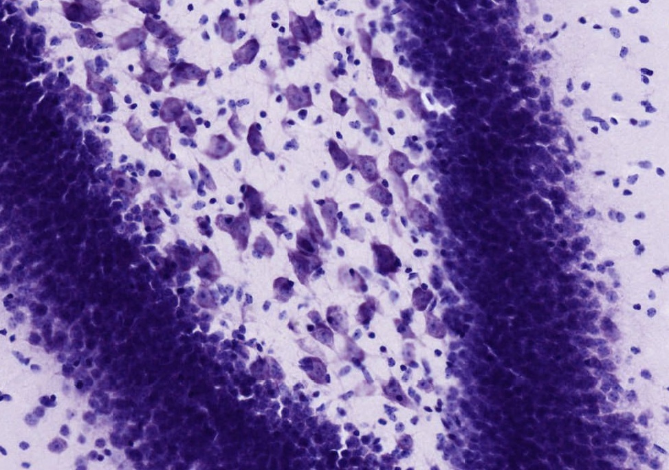
 Motor nerve cell from ventral horn of medulla spinalis of rabbit. The angular ande spindle-shaped Nissl bodies are well shown.
A Nissl body (or Nissl granule or tigroid body) is a large granular body found in neurons. These granules are rough endoplasmic reticulum (with free ribosomes) and are the site of protein synthesis. It was named after Franz Nissl, German neurologist (1860-1919)[1].
Nissl bodies can be demonstrated by a method of selective staining developed by Nissl (Nissl staining), using an aniline stain to label extranuclear RNA granules. This staining method is useful to localize the perikaryon, cell body, as it can be seen in the Soma (biology) and dendrites of neurons, though not in the axon or axon hillock. Due to RNA's basophilic (lat. "base-loving") properties it is stained blue by this method.
Nissl bodies show changes under various physiological conditions and in pathological conditions they may dissolve and disappear (karyolysis).
They are thought to be involved in the synthesis of neurotransmitters such as acetylcholine.
See also
References
External links
- MeSH Nissl+Bodies
- Histology at BU 04103loa - "Nervous Tissue and Neuromuscular Junction: spinal cord, cell bodies of anterior horn cells"
- Anatomy at MUN nerve/nerve97 (halfway down page)
- Histology at anhb.uwa.edu.au
- Tissues containing Nissl bodies at harvard.edu
Histology: nervous tissue (TA A14, GA 9.849, TH H2.00.06, H3.11) CNS GeneralGrey matter · White matter (Projection fibers · Association fiber · Commissural fiber · Lemniscus · Funiculus · Fasciculus · Decussation · Commissure) · meningesOtherPNS GeneralPosterior (Root, Ganglion, Ramus) · Anterior (Root, Ramus) · rami communicantes (Gray, White) · Autonomic ganglion (Preganglionic nerve fibers · Postganglionic nerve fibers)Myelination: Schwann cell (Neurolemma, Myelin incisure, Myelin sheath gap, Internodal segment)
Satellite glial cellNeurons/
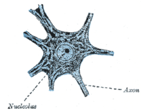
nerve fibersPartsPerikaryon (Axon hillock)
Axon (Axon terminals, Axoplasm, Axolemma, Neurofibril/neurofilament)
Dendrite (Nissl body, Dendritic spine, Apical dendrite/Basal dendrite)TypesGSA · GVA · SSA · SVA
fibers (Ia, Ib or Golgi, II or Aβ, III or Aδ or fast pain, IV or C or slow pain)GSE · GVE · SVE
Upper motor neuron · Lower motor neuron (α motorneuron, γ motorneuron, β motorneuron)Termination SynapseCategories:- Nervous tissue cells
- Neuroscience stubs

Wikimedia Foundation. 2010.