- Schwann cell
-
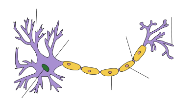
Structure of a typical neuron Schwann cells wrapped around an axon Schwann cells or neurolemmocytes are the principal glia of the peripheral nervous system (PNS). Glial cells function to support neurons and in the PNS, also include satellite cells, olfactory ensheathing cells, enteric glia and glia that reside at sensory nerve endings, such as the Pacinian corpuscle. Myelinating Schwann cells wrap around axons of motor and sensory neurons to form the myelin sheath.
Schwann cells are involved in many important aspects of peripheral nerve biology - the conduction of nervous impulses along axons, nerve development and regeneration, trophic support for neurons, production of the nerve extracellular matrix, modulation of neuromuscular synaptic activity, and presentation of antigens to T-lymphocytes. Charcot-Marie-Tooth disease (CMT), Guillain-Barré syndrome (GBS), schwannomatosis and chronic inflammatory demyelinating polyneuropathy (CIDP) are all neuropathies involving Schwann cells.
Contents
Description
 A Schwann cell in culture.
Named after the German physiologist Theodor Schwann, Schwann cells are a variety of glial cell that keep peripheral nerve fibres (both myelinated and unmyelinated) alive. In myelinated axons, Schwann cells form the myelin sheath (see above). The sheath is not continuous. Individual myelinating Schwann cells cover about 100 micrometres of an axon - equating to approximately 10,000 Schwann cells along a 1 metre length of the axon - which can be up to a metre or more in length. The gaps between adjacent Schwann cells are called nodes of Ranvier (see above). The vertebrate nervous system relies on the myelin sheath for insulation and as a method of decreasing membrane capacitance in the axon. The action potential jumps from node to node, in a process called saltatory conduction, which can increase conduction velocity up to ten times, without an increase in axonal diameter. In this sense, Schwann cells are the peripheral nervous system's analogues of the central nervous system's oligodendrocytes. However, unlike oligodendrocytes, each myelinating Schwann cell provides insulation to only one axon (see image). This arrangement permits saltatory conduction of action potentials with repropagation at the nodes of Ranvier. In this way, myelination greatly increases speed of conduction and saves energy.[1]
Non-myelinating Schwann cells are involved in maintenance of axons and are crucial for neuronal survival. Some group around smaller axons (External image here) and form Remak bundles.[2]
Myelinating Schwann cells begin to form the myelin sheath in mammals during fetal development and work by spiraling around the axon, sometimes with as many as 100 revolutions. A well-developed Schwann cell is shaped like a rolled-up sheet of paper, with layers of myelin in between each coil. The inner layers of the wrapping, which are predominantly membrane material, form the myelin sheath while the outermost layer of nucleated cytoplasm forms the neurolemma. Only a small volume of residual cytoplasm communicates the inner from the outer layers. This is seen histologically as the Schmidt-Lantermann incisure.
A number of experimental studies since 2001 have implanted Schwann cells in an attempt to induce remyelination in multiple sclerosis-afflicted patients.[3] In the past two decades, many studies have demonstrated positive results and potential for Schwann cell transplantation as a therapy for spinal cord injury, both in aiding regrowth and myelination of damaged CNS axons.[4] Indeed, Schwann cells are known for their roles in supporting nerve regeneration.[5] Nerves in the PNS consist of many axons myelinated by Schwann cells. If damage occurs to a nerve, the Schwann cells will aid in digestion of its axons (phagocytosis). Following this process, the Schwann cells can guide regeneration by forming a type of tunnel that leads toward the target neurons. The stump of the damaged axon is able to sprout, and those sprouts that grow through the Schwann-cell “tunnel” do so at the rate of approximately 1mm/day in good conditions. The rate of regeneration decreases with time. Successful axons can therefore reconnect with the muscles or organs they previously controlled with the help of Schwann cells, however, specificity is not maintained and errors are frequent, especially when long distances are involved.[6] If Schwann cells are prevented from associating with axons, the axons die. Regenerating axons will not reach any target unless Schwann cells are there to support them and guide them. They have been shown to be in advance of the growth cones. Schwann cells are essential for the maintenance of healthy axons. They produce a variety of factors, including neurotrophins, and also transfer essential molecules across to axons.
Schwann cell lineage
Schwann cells are of neural crest origin. During mouse embryonic development, neural crest cells first differentiate into Schwann cell precursors (SCPs) at around embryonic day (E) 12-13. These precursor cells subsequently differentiate into immature Schwann cells at approximately E15-16, persisting until birth. The postnatal fate of the immature Schwann cell depends on its random association with axons. In a process called radial sorting, whereby Schwann cells segregate axons by extending processes into axon bundles, the Schwann cells that happen to associate with a large diameter axon (>1 μm) will develop into myelinating Schwann cells. Small diameter axons become entrenched in the invaginations of non-myelinating Schwann cells, also called Remak bundles. A key regulator of this process is the axonally-derived signal Neuregulin-1, which binds to cell surface receptors on the Schwann cell and promotes myelination of large diameter axons and sorting of small diameter axons in Remak bundles, dependent on the activity of the β-secretase BACE1 ([7][8][9][10][11]). A further class of non-myelinating Schwann cell, the terminal (or perisynaptic) Schwann cell, exists at the neuromuscular junction, in close proximity to the neuron-muscle synapse. The transition from immature Schwann cell to myelinating/non-myelinating Schwann cell is reversible. When the nerve is injured, Schwann cells can dedifferentiate to form a cell type resembling the immature Schwann cell, often referred to as a denervated or dedifferentiated Schwann cell. This allows them to re-enter the cell cycle in order to proliferate and aid nerve regeneration.[12]
Immunoreactivity
The different classes of Schwann cells express characteristic antigenic markers that can be targeted with antibodies. Myelinating Schwann cells can be visualised by immunohistochemistry using antibodies against the proteins S-100, Myelin protein zero (P-Zero) and Myelin basic protein (MBP). Non-myelinating Schwann cells such as those that form Remak bundles and terminal Schwann cells are positive for S-100 and Glial fibrillary acidic protein (GFAP).
See also
- Electrophysiology
- Hodgkin-Huxley Model
- Neurotransmission
- Schwannoma
References
- ^ Kalat, James W. Biological Psychology, 9th ed. USA: Thompson Learning, 2007.[page needed]
- ^ "Medical Student Named Rhodes Scholar" - FOCUS - News from Harvard Medical, Dental and Public Health Schools - Copyright © 2010 President and Fellows of Harvard College.
- ^ "First surgical transplant attempted to repair myelin - News - Brief Article". Inside MS. 2001. http://www.findarticles.com/p/articles/mi_m0850/is_4_19/ai_79957646.
- ^ Oudega, Martin; Xu, Xiao-Ming (2006). "Schwann Cell Transplantation for Repair of the Adult Spinal Cord". Journal of Neurotrauma 23 (3–4): 453–67. doi:10.1089/neu.2006.23.453. PMID 16629629.
- ^ Bhatheja, Kanav; Field, Jeffrey (2006). "Schwann cells: Origins and role in axonal maintenance and regeneration". The International Journal of Biochemistry & Cell Biology 38 (12): 1995–9. doi:10.1016/j.biocel.2006.05.007.
- ^ Carlson, Neil R. Physiology of Behavior, 9th ed. USA: Pearson Education, Inc., 2007.[page needed]
- ^ Garratt, A. N.; Voiculescu, O; Topilko, P; Charnay, P; Birchmeier, C (2000). "A Dual Role of erbB2 in Myelination and in Expansion of the Schwann Cell Precursor Pool". The Journal of Cell Biology 148 (5): 1035–46. doi:10.1083/jcb.148.5.1035. PMC 2174554. PMID 10704452. http://www.pubmedcentral.nih.gov/articlerender.fcgi?tool=pmcentrez&artid=2174554.
- ^ Michailov, G. V.; Sereda, MW; Brinkmann, BG; Fischer, TM; Haug, B; Birchmeier, C; Role, L; Lai, C et al. (2004). "Axonal Neuregulin-1 Regulates Myelin Sheath Thickness". Science 304 (5671): 700–3. doi:10.1126/science.1095862. PMID 15044753.
- ^ Taveggia, Carla; Zanazzi, George; Petrylak, Ashley; Yano, Hiroko; Rosenbluth, Jack; Einheber, Steven; Xu, Xiaorong; Esper, Raymond M. et al. (2005). "Neuregulin-1 Type III Determines the Ensheathment Fate of Axons". Neuron 47 (5): 681–94. doi:10.1016/j.neuron.2005.08.017. PMC 2387056. PMID 16129398. http://www.pubmedcentral.nih.gov/articlerender.fcgi?tool=pmcentrez&artid=2387056.
- ^ Willem, M.; Garratt, A. N.; Novak, B.; Citron, M.; Kaufmann, S.; Rittger, A.; Destrooper, B.; Saftig, P. et al. (2006). "Control of Peripheral Nerve Myelination by the -Secretase BACE1". Science 314 (5799): 664–6. doi:10.1126/science.1132341. PMID 16990514.
- ^ Hu, Xiangyou; Hicks, Caitlin W; He, Wanxia; Wong, Philip; MacKlin, Wendy B; Trapp, Bruce D; Yan, Riqiang (2006). "Bace1 modulates myelination in the central and peripheral nervous system". Nature Neuroscience 9 (12): 1520–5. doi:10.1038/nn1797. PMID 17099708.
- ^ Jessen, Kristjan R.; Mirsky, Rhona (2005). "The origin and development of glial cells in peripheral nerves". Nature Reviews Neuroscience 6 (9): 671–682. doi:10.1038/nrn1746. PMID 16136171.
External links
- Diagram at clc.uc.edu
- Histology at BU 21301loa - "Ultrastructure of the Cell: myelinated axon and Schwann cell"
- Cell Centered Database - Schwann cell
Histology: nervous tissue (TA A14, GA 9.849, TH H2.00.06, H3.11) CNS GeneralGrey matter · White matter (Projection fibers · Association fiber · Commissural fiber · Lemniscus · Funiculus · Fasciculus · Decussation · Commissure) · meningesOtherPNS GeneralPosterior (Root, Ganglion, Ramus) · Anterior (Root, Ramus) · rami communicantes (Gray, White) · Autonomic ganglion (Preganglionic nerve fibers · Postganglionic nerve fibers)Myelination: Schwann cell (Neurolemma, Myelin incisure, Myelin sheath gap, Internodal segment)
Satellite glial cellNeurons/
nerve fibersPartsPerikaryon (Axon hillock)
Axon (Axon terminals, Axoplasm, Axolemma, Neurofibril/neurofilament)
Dendrite (Nissl body, Dendritic spine, Apical dendrite/Basal dendrite)TypesGSA · GVA · SSA · SVA
fibers (Ia, Ib or Golgi, II or Aβ, III or Aδ or fast pain, IV or C or slow pain)GSE · GVE · SVE
Upper motor neuron · Lower motor neuron (α motorneuron, γ motorneuron, β motorneuron)Termination SynapseHuman cell types / list derived primarily from ectoderm Surface ectoderm Trichocyte · KeratinocyteNeural crest glia: Schwann cell · Satellite glial cellDigestive systemNeural tube Categories:- Neurophysiology
- Glial cells
Wikimedia Foundation. 2010.

