- Cardiac plexus
Infobox Nerve
Name = PAGENAME
Latin = plexus cardiacus
GraySubject = 220
GrayPage = 984
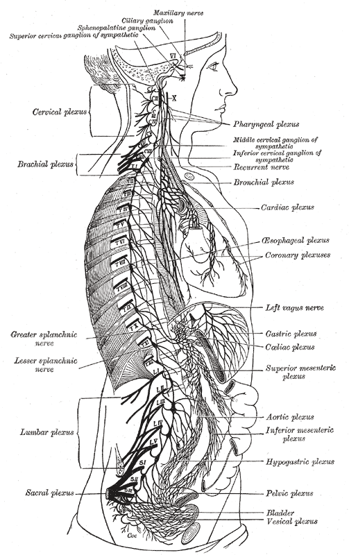
Caption = The right sympathetic chain and its connections with the thoracic, abdominal, and pelvic plexuses. (Cardiac plexus labeled at center right.)
Caption2 =
Innervates =
BranchFrom =
BranchTo =
MeshName =
MeshNumber =
DorlandsPre = p_24
DorlandsSuf = 12647601
The cardiac plexus is aplexus of nerves situated at the base of theheart that innervate the heart.tructure
The cardiac plexus is divided into a superficial part, which lies in the concavity of the
aortic arch , and a deep part, between the aortic arch and the trachea.The two parts are, however, closely connected.
uperficial part
The superficial part of the cardiac plexus lies beneath the arch of the aorta, in front of the
right pulmonary artery .It is formed by the superior cardiac branch of the left
sympathetic trunk and the lower superior cervical cardiac branch of the leftvagus nerve .A small ganglion, the "cardiac ganglion of Wrisberg", is occasionally found connected with these nerves at their point of junction.
This ganglion, when present, is situated immediately beneath the arch of the aorta, on the right side of the
ligamentum arteriosum .The superficial part of the cardiac plexus gives branches
* (a) to the deep part of the plexus;
* (b) to the anterior coronary plexus; and
* (c) to the left anteriorpulmonary plexus .Deep part
The deep part of the cardiac plexus is situated in front of the bifurcation of the trachea, above the point of division of the pulmonary artery, and behind the aortic arch.
It is formed by the cardiac nerves derived from the cervical ganglia of the sympathetic trunk, and the cardiac branches of the vagus and
recurrent laryngeal nerve s.The only cardiac nerves which do not enter into the formation of the deep part of the cardiac plexus are the superior cardiac nerve of the left sympathetic trunk, and the lower of the two superior cervical cardiac branches from the left vagus nerve, which pass to the superficial part of the plexus.
Right half
The branches from the right half of the deep part of the cardiac plexus pass, some in front of, and others behind, the right pulmonary artery; the former, the more numerous, transmit a few filaments to the anterior pulmonary plexus, and are then continued onward to form part of the anterior coronary plexus; those behind the pulmonary artery distribute a few filaments to the right atrium, and are then continued onward to form part of the posterior coronary plexus.
Left half
The left half of the deep part of the plexus is connected with the superficial part of the cardiac plexus, and gives filaments to the left atrium, and to the anterior pulmonary plexus, and is then continued to form the greater part of the posterior coronary plexus.
ee also
*
Sympathetic trunk
*Splanchnic nerves
*Autonomic nervous system External links
* (superficial)
* (deep)
* (NormanAnatomyFig|thoraxautonomicner)
*
Wikimedia Foundation. 2010.