- Locus coeruleus
-
Brain: Locus coeruleus 
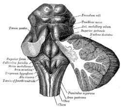
Rhomboid fossa. (Locus coeruleus not labeled, but is very near [just lateral to] colliculus facialis, which is labeled at center left.) 
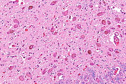
Micrograph showing the locus coeruleus. HE-LFB stain. Latin locus caeruleus Gray's subject #187 778 NeuroNames hier-578 NeuroLex ID birnlex_905 The locus coeruleus (also spelled locus caeruleus or locus ceruleus), is a nucleus in the brainstem involved with physiological responses to stress and panic. It was discovered in the 18th century by Félix Vicq-d'Azyr.
The locus coeruleus is the principal site for brain synthesis of norepinephrine (noradrenaline). The locus coeruleus and the areas of the body affected by the norepinephrine it produces are described collectively as the locus coeruleus-noradrenergic system or LC-NA system.[1] Norepinephrine may also be released directly into the blood from the adrenal medulla.
The name is derived from the Latin words coeruleus and locus. Literally, this means "the blue spot", a name derived from its azure appearance in unstained brain tissue. The color is due to light scattering from melanin in noradrenergic (producing or activated by norepinephrine) nerve cell bodies. Caeruleus is the classical Latin spelling, but coeruleus is the more common spelling. The spelling ceruleus, formed by contraction of the digraph ae or oe into e, is an American English form.
Contents
Anatomy
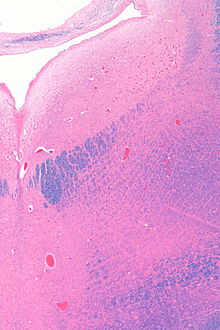
 Micrograph showing the locus coeruleus (upper-right of image) in an axial section of the pons. The fourth ventricle (quasi-triangular white area) is in the upper left of the image. The midline is seen on the left. The large white area in the upper-left corner is where the cerebellum would be. HE-LFB stain.
The locus coeruleus (or "LC") is in the dorsal wall of the rostral pons in the lateral floor of the fourth ventricle. It is composed of mostly medium-size neurons. Melanin granules inside the neurons of the LC contribute to its blue color. Thus, it is also known as the nucleus pigmentosus pontis, meaning "heavily pigmented nucleus of the pons." The neuromelanin is formed by the polymerization of noradrenaline and is analogous to the black dopamine-based neuromelanin in the substantia nigra.
In adult humans (19-78) the locus coeruleus has 22,000 to 51,000 total pigmented neurons that range in size between 31,000 to 60, 000 μm3.[2]
Connections
The projections of this nucleus reach far and wide. For example, they innervate the spinal cord, the brain stem, cerebellum, hypothalamus, the thalamic relay nuclei, the amygdala, the basal telencephalon, and the cortex. The norepinephrine from the LC has an excitatory effect on most of the brain, mediating arousal and priming the brain’s neurons to be activated by stimuli.
As an important homeostatic control center of the body, the locus coeruleus receives afferents from the hypothalamus. The cingulate gyrus and the amygdala also innervate the LC, allowing emotional pain and stressors to trigger noradrenergic responses. The cerebellum and afferents from the raphe nuclei also project to the LC, particularly the raphe pontis and raphe dorsalis.
The locus coeruleus receives inputs from a number of other brain regions, primarily:
- Medial prefrontal cortex, whose connection is constant, excitatory, and increases in strength with raised activity levels in the subject
- Nucleus paragigantocellularis, which integrates autonomic and environmental stimuli
- Nucleus prepositus hypoglossi, which is involved in gaze
- Lateral hypothalamus, which releases orexin, which, as well as its other functions, is excitatory in the locus coeruleus.
Function
The locus coeruleus may figure in clinical depression, panic disorder, and anxiety. Some medications including norepinephrine reuptake inhibitors (reboxetine, atomoxetine), serotonin-norepinephrine reuptake inhibitors (venlafaxine), and norepinephrine-dopamine reuptake inhibitors (bupropion) are believed to show efficacy by acting upon neurons in this area. This area of the brain is also intimately involved in REM sleep.
Psychiatric research has documented that enhanced noradrenergic postsynaptic responsiveness in the neuronal pathway (brain circuit) that originates in the locus coeruleus and end in the basolateral nucleus of the amygdala is a major factor in the pathophysiology of most stress-induced fear-circuitry disorders and especially in posttraumatic stress disorder (PTSD). The LC neurons are probably the origin of the first or second “leg” of what has been recently termed the "PTSD candidate circuit." Combat-related PTSD (in a 2005 study of deceased American army veterans from World War II) was shown to be associated with a postmortem diminished number of neurons in the locus coeruleus (LC) on the right side of the brain.[3] The role of the LC in PTSD may explain the dramatic effectiveness of two generic medications, propranolol and prazosin for the secondary prevention and treatment of PTSD, respectively.[citation needed]
In stress
The locus coeruleus is responsible for mediating many of the sympathetic effects during stress. The locus coeruleus is activated by stress, and will respond by increasing norepinephrine secretion, which in turn will alter cognitive function (through the prefrontal cortex), increase motivation (through nucleus accumbens), activate the hypothalamic-pituitary-adrenal axis, and increase the sympathetic discharge/inhibit parasympathetic tone (through the brainstem). Specific to the activation of the hypothalamo-pituitary adrenal axis, norepinephrine will stimulate the secretion of corticotropin-releasing factor from the hypothalamus, which induces adrenocorticotropic hormone release from the anterior pituitary and subsequent cortisol synthesis in the adrenal glands. Norepinephrine released from locus coeruleus will feedback to inhibit its production, and corticotropin-releasing hormone will feedback to inhibit its production, while positively feeding to the locus coeruleus to increase norepinephrine production.[4]
The LC's role in cognitive function in relation to stress is complex and multi-modal. Norepinephrine released from the LC can act on α2 receptors to increase working memory, or an excess of NE may decrease working memory by binding to the lower affinity α1 receptors.[5]
In opiate withdrawal
Opioids inhibit the firing of neurons in the locus coeruleus. When opioid consumption is stopped, the increased activity of the locus coeruleus contributes to the symptoms of opiate withdrawal. The alpha2 adrenoceptor agonist clonidine is used to counteract this withdrawal effect by decreasing adrenergic neurotransmission from the locus coeruleus. [6]
Rett syndrome
The genetic defect of the transcriptional regulator MECP2 is responsible for Rett syndrome.[7] A MECP2 deficiency has been associated to catecholaminergic dysfunctions related to autonomic and sympathoadrenergic system in mouse models of Rett Syndrome (RTT). The Locus Coeruleus is the major source of noradrenergic innervation in the brain and sends widespread connections to rostral (cerebral cortex, hippocampus, hypothalamus) and caudal (cerebellum, brainstem nuclei) brain areas[8] and.[9] Indeed, an alteration of this structure could contribute to several symptoms observed in MECP2-deficient mice. Changes in the electrophysiological properties of cells in the locus ceruleus were shown. These Locus Coeruleus cell changes include hyperexcitability and decreased functioning of its noradrenergic innervation.[10] Interestingly, a reduction of the tyrosine hydroxylase (TH) mRNA level, the rate-limiting enzyme in catecholamine synthesis, was detected in the whole pons of MECP2-null male as well as in adult heterozygous female mice. Using immunoquantification techniques, a decrease of TH protein staining level, number of locus coeruleus TH-expressing neurons and density of dendritic arborization surrounding the structure was shown in symptomatic MECP2-deficient mice.[11] However, locus coeruleus cells are not dying but are more likely losing their fully mature phenotype since no apoptotic neurons in the pons were detected.[11] Researchers have concluded that "Because these neurons are a pivotal source of norepinephrine throughout the brainstem and forebrain and are involved in the regulation of diverse functions disrupted in Rett Syndrome, such as respiration and cognition, we hypothesize that the locus ceruleus is a critical site at which loss of MECP2 results in CNS dysfunction. Restoration of normal locus ceruleus function may therefore be of potential therapeutic value in the treatment of Rett Syndrome.[10] This could explain why a norepinephrine reuptake inhibitor (desipramine, DMI) which enhances the extracellular NE levels at all noradrenergic synapses ameliorated some Rett syndrome symptoms in a mouse model of Rett syndrome.[11]
Alzheimer's disease
There is up to 70% loss of locus ceruleus neurons in Alzheimer's disease.[12] Mouse models of Alzheimer's disease show accelerated progression after chemical destruction of the locus ceruleus [13] The norepinephrine from locus ceruleus cells in addition to its neurotransmitter role locally defuses from "varicosities". As such it provides an endogenous antiinflammatory agent in the microenvironment around the neurons, glial cells, and blood vessels in the neocortex and hippocampus.[14] It has been shown that norepinephrine stimulates mouse microglia to suppress Aβ-induced production of cytokines and their phagocytosis of Aβ.[14] This suggests that degeneration of the locus ceruleus might be responsible for increased Aβ deposition in AD brains.[14]
See also
- Raphe nucleus
- Substantia nigra
- Reticular formation
References
- ^ Mehler, Mark F.; Dominick P. Purpura (March 2009). "Autism, fever, epigenetics and the locus coeruleus". Brain Research Reviews 59 (2): 388–392. doi:10.1016/j.brainresrev.2008.11.001. PMC 2668953. PMID 19059284. http://www.sciencedirect.com/science/article/pii/S0165017308001379. Retrieved 9 June 2011.
- ^ Mouton PR, Pakkenberg B, Gundersen HJ, Price DL (August 1994). "Absolute number and size of pigmented locus coeruleus neurons in young and aged individuals". J. Chem. Neuroanat. 7 (3): 185–90. PMID 7848573.
- ^ Bracha HS, Garcia-Rill E, Mrak RE, Skinner R (2005). "Postmortem locus coeruleus neuron count in three American veterans with probable or possible war-related PTSD". The Journal of neuropsychiatry and clinical neurosciences 17 (4): 503–9. doi:10.1176/appi.neuropsych.17.4.503. PMID 16387990.
- ^ Benarroch EE. The locus ceruleus norepinephrine system: functional organization and potential clinical significance. Neurology. 2009 Nov 17;73(20):1699-704.
- ^ Ramos BP, Arnsten AF. Adrenergic pharmacology and cognition: focus on the prefrontal cortex. Pharmacol Ther 2007; 113: 523-536.
- ^ P. Devenyi, A. Mitwalli, and W. Graham Clonidine therapy for narcotic withdrawal. Can Med Assoc 1982 November 15; 127(10): 1009–1011. http://www.ncbi.nlm.nih.gov/pmc/articles/PMC1862300/?page=1
- ^ Amir RE, Van den Veyver IB, Wan M, Tran CQ, Francke U, Zoghbi HY (1999) Rett Syndrome is caused by mutations in X-linked MECP2, encoding methyl-CpG-binding protein 2 Nat Genet. 1999 Oct 23(2):185-8
- ^ Hokfelt T,Martensson R,Bjorklund A,Kleinau S,Goldstein M. 1984. Distribution maps of tyrosine-hydroxylase-immunoreactive neurons in the rat brain. In Handbook of Chemical Neuroanatomy, Vol. 2. Classical Transmitters in the CNS, Part I ( A. Bjorklund and T. Hokfelt, eds.) pp. 277-379. Elsevier, New York.
- ^ Berridge CW,Waterhouse BD 2003 The locus coeruleus-noradrenergic system: modulation of behavioral state and state-dependent cognitive processes. Brain Res Rev 42: 33-84
- ^ a b Taneja P, Ogier M, Brooks-Harris G, Schmid DA, Katz DM, Nelson SB (2009). "Pathophysiology of Locus Ceruleus Neurons in a Mouse Model of Rett Syndrome". Journal of Neuroscience 29 (39): 12187–12195. doi:10.1523/JNEUROSCI.3156-09.2009.
- ^ a b c Roux JC, Panayotis N, Dura E, Villard L. (2009) Progressive Noradrenergic Deficits in the Locus Coeruleus of MECP2 Deficient Mice J Neurosci Res http://www3.interscience.wiley.com/cgi-bin/fulltext/123208150/HTMLSTART
- ^ Bondareff W, Mountjoy CQ, Roth M. Loss of neurons of origin of the adrenergic projection to cerebral cortex (nucleus locus ceruleus) in senile dementia. Neurology. 1982 Feb;32(2):164-8.
- ^ Heneka MT, Ramanathan M, Jacobs AH, Dumitrescu-Ozimek L, Bilkei-Gorzo A, Debeir T, Sastre M, Galldiks N, Zimmer A, Hoehn M, Heiss WD, Klockgether T, Staufenbiel M. Locus ceruleus degeneration promotes Alzheimer pathogenesis in amyloid precursor protein 23 transgenic mice. J Neurosci. 2006 Feb 1;26(5):1343-54.
- ^ a b c Heneka MT, Nadrigny F, Regen T, Martinez-Hernandez A, Dumitrescu-Ozimek L, Terwel D, Jardanhazi-Kurutz D, Walter J, Kirchhoff F, Hanisch UK, Kummer MP. (2010). Locus ceruleus controls Alzheimer's disease pathology by modulating microglial functions through norepinephrine. Proc Natl Acad Sci U S A. 107:6058–6063 doi:10.1073/pnas.0909586107 PMID 20231476
External links
- "A Lecture, Higher Brain Function: Activation of the Brain and Levels of Consciousness" at East Tennessee State University
- BrainMaps at UCDavis locus coeruleus
- Diagram at University of Texas at Austin
- Diagram at University of Virginia
- http://www2.umdnj.edu/~neuro/studyaid/Practical2000/Q45.htm
Human brain, rhombencephalon, metencephalon: pons (TA A14.1.05.101–604, GA 9.785) Dorsal/
(tegmentum)SurfaceWhite: Sensory/ascendingTrapezoid body/VIII · Trigeminal lemniscus (Dorsal trigeminal tract, Ventral trigeminal tract) · Medial lemniscus · Lateral lemniscus
MLF, III, IV and VI: Vestibulo-oculomotor fibers
Anterior trigeminothalamic tract · Central tegmental tractWhite: Motor/descendingICP (Vestibulocerebellar tract)
MLF, III, IV and VI: Vestibulospinal tract (Medial vestibulospinal tract, Lateral vestibulospinal tract)Other greyApneustic center · Pneumotaxic center (Medial parabrachial nucleus) · Lateral parabrachial nucleus · Superior olivary nucleus · Caerulean nucleusVentral/
(base)White: Motor/descendingSurfaceBasilar sulcusOther grey: Raphe/
reticularVentricular system, rhombencephalon, met- and myel-: fourth ventricle (TA A14.1.05.701–726, GA 9.797) Roof (dorsal) rostral: Superior medullary velum (Frenulum)
caudal: Inferior medullary velum · Taenia of fourth ventricleFloor/rhomboid fossa (ventral) rostral (pons): Facial colliculus · Locus coeruleus
caudal (medulla}: Vagal trigone · Hypoglossal trigone · Area postrema · Obex
Medial eminence · Sulcus limitansApertures Other Tela chorioidea of fourth ventricle · FastigiumNeurotransmitter systems Acetylcholine BA/M Mesocortical pathway: Ventral tegmental area → Frontal cortex
Mesolimbic pathway: Ventral tegmental area → Nucleus accumbens
Nigrostriatal pathway: Pars compacta → Striatum
Tuberoinfundibular pathway: Hypothalamus → Pituitary glandLocus coeruleusSerotonin pathwaysAA Categories:- Brainstem
- Neuroanatomy
Wikimedia Foundation. 2010.
