- Taenia of fourth ventricle
Infobox Brain
Name = PAGENAME
Latin = taenia ventriculi quarti
GraySubject = 187
GrayPage = 797

Caption =Rhomboid fossa . (Taenia of fourth ventricle labeled at bottom left.)
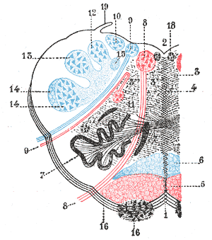
Caption2 = Theformatio reticularis of themedulla oblongata , shown by a transverse section passing through the middle of theolive . (Testut.) 1.Anterior median fissure . 2.Fourth ventricle . 3.Formatio reticularis , with 3’, its internal part (reticularis alba), and 3’’, its external part (reticularis grisea). 4.Raphé . 5.Pyramid . 6.Lemniscus . 7.Inferior olivary nucleus with the twoaccessory olivary nuclei . 8.Hypoglossal nerve , with 8’, its nucleus of origin. 9.Vagus nerve , with 9’, its nucleus of termination. 10.Lateral dorsal acoustic nucleus . 11.Nucleus ambiguus (nucleus of origin of motor fibers ofglossopharyngeal ,vagus , and cerebral portion ofspinal accessory ). 12.Gracile nucleus . 13.Cuneate nucleus . 14. Head ofposterior column , with 14’, the lower sensory root oftrigeminal nerve . 15.Fasciculus solitarius . 16.Anterior external arcuate fibers , with 16’, thenucleus arcuatus . 17.Nucleus lateralis 18. Nucleus offasciculus teres . 19.Ligula .
IsPartOf =
Components =
Artery =
Vein =
Acronym =
BrainInfoType = hier
BrainInfoNumber = 629
MeshName =
MeshNumber =
DorlandsPre = t_01
DorlandsSuf = 12789539
The taenia of the fourth ventricle (ligula, tenia of fourth ventricle) are two narrow bands ofwhite matter , one on either side, which complete the lower part of the roof of the cavity.Each consists of a vertical and a horizontal part.
* The "vertical part" is continuous below the
obex with theclava , to which it is adherent by its lateral border.* The "horizontal portion" extends transversely across the
inferior peduncle , below thestriæ medullares , and roofs in the lower and posterior part of thelateral recess ; it is attached by its lower margin to the inferior peduncle, and partly encloses thechoroid plexus , which, however, projects beyond it like a cluster of grapes; and hence this part of the tænia has been termed the "cornucopia".ee also
*
fourth ventricle
=Additional
Wikimedia Foundation. 2010.