- Inner ear
-
Inner ear 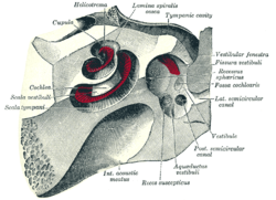
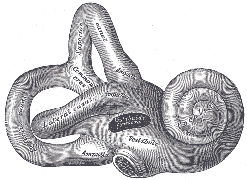
The cochlea and vestibule, viewed from above. Latin auris interna Gray's subject #232 1047 Artery labyrinthine artery MeSH Inner+ear 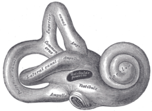 Inner ear
The inner ear is the innermost part of the vertebrate ear. In mammals, it consists of the bony labyrinth, a system of passages comprising two main functional parts:
- The cochlea, dedicated to hearing
- The vestibular system, dedicated to balance
The inner ear is found in all vertebrates, with substantial variations in form and function. The inner ear is innervated by the eighth cranial nerve in all vertebrates.
Contents
Embryology
The human inner ear develops during week 4 of embryonic development from the auditory placode, a thickening of the ectoderm which gives rise to the bipolar neurons of the cochlear and vestibular ganglions.[1] As the auditory placode invaginates towards the embryonic mesoderm, it forms the auditory vesicle or otocysts.
The auditory vesicle will give rise to the utricluar and saccular components of the membranous labyrinth. They contain the sensory hair cells and otoliths of the macula of utricle and of the saccule, respectively, which respond to linear acceleration and the force of gravity. The utricular division of the auditory vesicle also responds to angular acceleration, as well as the endolymphatic sac and duct that connect the saccule and utricle.
Beginning in the fifth week of development, the auditory vesicle also gives rise to the cochlear duct, which contains the spiral organ of Corti and the endolymph that accumulates in the membranous labyrinth.[2] The vestibular wall will separate the cochlear duct from the perilymphatic scala vestibuli, a cavity inside the cochlea. The basilar membrane separates the cochlear duct from the scala tympani, a cavity within the cochlear labyrinth. The lateral wall of the cochlear duct is formed by the spiral ligament and the stria vascularis, which produces the endolymph. The hair cells develop from the lateral and medial ridges of the cochlear duct, which together with the tectorial membrane make up the organ of Corti.[2]
Divisions of labyrinth
The labyrinth can be divided by layer or by region.
Bony vs. membranous
The bony labyrinth, or osseous labyrinth, is the network of passages with bony walls lined with periosteum. The membranous labyrinth runs inside of the bony labryinth. There is a layer of perilymph fluid between them. The three parts of the bony labyrinth are the vestibule of the ear, the semicircular canals, and the cochlea.
Vestibular vs. cochlear
In the middle ear, the energy of pressure waves is translated into mechanical vibrations. The cochlea propagates these mechanical signals as waves in fluid and membranes, and finally transduces them to nerve impulses which are transmitted to the brain.[3]
The vestibular system is the region of the inner ear where the semicircular canals converge, close to the cochlea. The vestibular system works with the visual system to keep objects in focus when the head is moving. Joint and muscle receptors also are important in maintaining balance. The brain receives, interprets, and processes the information from these systems to control balance.
The vestibular system of the inner ear is responsible for the sensations of balance and motion. It uses the same kinds of fluids and detection cells (hair cells) as the cochlea uses, and sends information to the brain about the attitude, rotation, and linear motion of the head. The type of motion or attitude detected by a hair cell depends on its associated mechanical structures, such as the curved tube of a semicircular canal or the calcium carbonate crystals (otolith) of the saccule and utricle.
Pathology
Interference with or infection of the labyrinth can result in a syndrome of ailments called labyrinthitis. The symptoms of Labyrinthitis include temporary nausea, disorientation, vertigo, and dizziness. Labyrinthitis can be caused by viral infections, bacterial infections, or physical blockage of the inner ear.[4][5]
Anatomical details
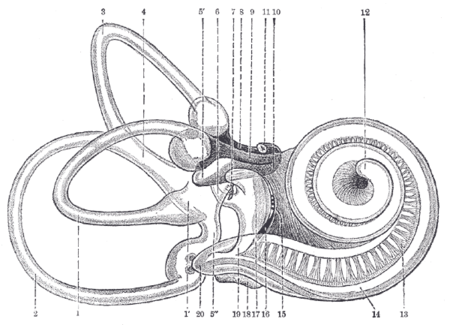
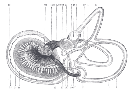
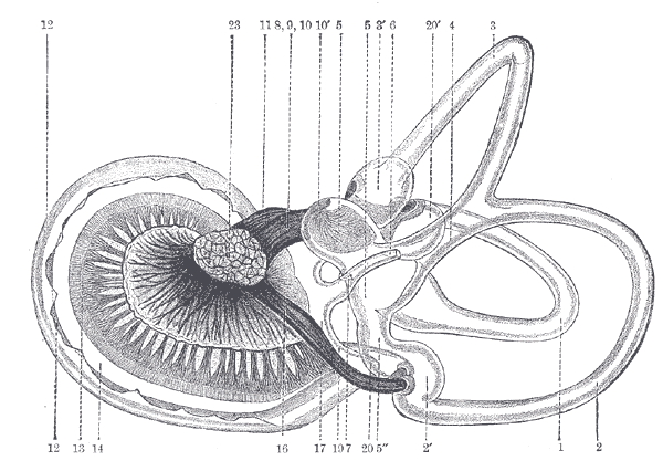
Top image is antero-lateral and bottom image is postero-medial.
- Lateral semicircular canal; 1’, its ampulla
- Posterior canal; 2’, its ampulla
- Superior canal; 3’, its ampulla
- Conjoined limb of superior and posterior canals (sinus utriculi superior)
- Utricle; 5’. Recessus utriculi; 5”. Sinus utriculi posterior
- Ductus endolymphaticus
- Canalis utriculosaccularis
- Nerve to ampulla of superior canal
- Nerve to ampulla of lateral canal
- Nerve to recessus utriculi (in top image, the three branches appear conjoined); 10’. Ending of nerve in recessus utriculi
- Facial nerve
- Lagena cochleæ
- Nerve of cochlea within spiral lamina
- Basilar membrane
- Nerve fibers to macula of saccule
- Nerve to ampulla of posterior canal
- Saccule
- Secondary membrane of tympanum
- Canalis reuniens
- Vestibular end of ductus cochlearis
- Section of the facial and acoustic nerves within internal acoustic meatus (the separation between them is not apparent in the section)
- (No entry)
- Vestibulocochlear nerve (auditory or acoustic, cranial nerve VIII)
Non-humans
Birds have an auditory system similar to that of mammals, including a cochlea. Reptiles, amphibians, and fish do not have cochleas but hear with simpler auditory organs or vestibular organs, which generally detect lower-frequency sounds than the cochlea.
The cochlear system
In reptiles, sound is transmitted to the inner ear by the stapes (stirrup) bone of the middle ear. This is pressed against the oval window, a membrane-covered opening on the surface of the vestibule. From here, sound waves are conducted through a short perilymphatic duct to a second opening, the round window, which equalizes pressure, allowing the incompressible fluid to move freely. Running parallel with the perilymphatic duct is a separate blind-ending duct, the lagena, filled with endolymph. The lagena is separated from the perilymphatic duct by a basilar membrane, and contains the sensory hair cells that finally translate the vibrations in the fluid into nerve signals. It is attached at one end to the saccule.[6]
In most reptiles the perilymphatic duct and lagena are relatively short, and the sensory cells are confined to a small basilar papilla lying between them. However, in birds, mammals, and crocodilians, these structures become much larger and somewhat more complicated. In birds, crocodilians, and monotremes, the ducts are simply extended, together forming an elongated, more or less straight, tube. The endolymphatic duct is wrapped in a simple loop around the lagena, with the basilar membrane lying along one side. The first half of the duct is now referred to as the scala vestibuli, while the second half, which includes the basilar membrane, is called the scala tympani. As a result of this increase in length, the basilar membrane and papilla are both extended, with the latter developing into the organ of Corti, while the lagena is now called the cochlear duct. All of these structures together constitute the cochlea.[6]
In mammals (other than monotremes), the cochlea is extended still further, becoming a coiled structure in order to accommodate its length within the head. The organ of Corti also has a more complex structure in mammals than it does in other amniotes.[6]
The arrangement of the inner ear in living amphibians is, in most respects, similar to that of reptiles. However, they often lack a basilar papilla, having instead an entirely separate set of sensory cells at the upper edge of the saccule, referred to as the papilla amphibiorum, which appear to have the same function.[6]
Although many fish are capable of hearing, the lagena is, at best, a short diverticulum of the saccule, and appears to have no role in sensation of sound. Various clusters of hair cells within the inner ear may instead be responsible; for example, bony fish contain a sensory cluster called the macula neglecta in the utricle that may have this function. Although fish have neither an outer nor a middle ear, sound may still be transmitted to the inner ear through the bones of the skull, or by the swim bladder, parts of which often lie close by in the body.[6]
The vestibular system
By comparison with the cochlear system, the vestibular system varies relatively little between the various groups of jawed vertebrates. The central part of the system consists of two chambers, the saccule and utricle, each of which includes one or two small clusters of sensory hair cells. All jawed vertebrates also possess three semicircular canals arising from the utricle, each with an ampulla containing sensory cells at one end.[6]
An endolymphatic duct runs from the saccule up through the head, and ending close to the brain. In cartilaginous fish, this duct actually opens onto the top of the head, and in some teleosts, it is simply blind-ending. In all other species, however, it ends in an endolymphatic sac. In many reptiles, fish, and amphibians this sac may reach considerable size. In amphibians the sacs from either side may fuse into a single structure, which often extends down the length of the body, parallel with the spinal canal.[6]
The primitive lampreys and hagfish, however, have a simpler system. The inner ear in these species consists of a single vestibular chamber, although in lampreys, this is associated with a series of sacs lined by cilia. Lampreys have only two semicircular canals, with the horizontal canal being absent, while hagfish have only a single, vertical, canal.[6]
Additional images
-
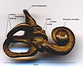
Ear labyrinth
-
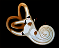
Inner ear
-
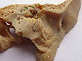
Temporal bone
-
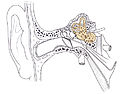
Internal ear
References
- ^ Hyman, Libbie Henrietta (1992). Hyman's comparative vertebrate anatomy (3 ed.). University of Chicago Press. p. 634. ISBN 0226870138. http://books.google.com/books?id=VKlWjdOkiMwC. Retrieved 2011-05-14.
- ^ a b Brauer, Philip R. (2003). Human embryology: the ultimate USMLE step 1 review. Elsevier Health Sciences. p. 61. ISBN 156053561X. http://books.google.com/books?id=_Cb_XXR5HCQC. Retrieved 2011-05-14.
- ^ Jan Schnupp, Israel Nelken and Andrew King (2011). Auditory Neuroscience. MIT Press. ISBN 026211318X. https://mustelid.physiol.ox.ac.uk/drupal/?q=ear.
- ^ "Labyrinthine Dysfunction During Diving". 1st Undersea and Hyperbaric Medical Society Workshop. (Undersea and Hyperbaric Medical Society) UHMS Publication Number WS6-15-74.: 11. 1973. http://archive.rubicon-foundation.org/4291. Retrieved 2009-03-11.
- ^ Kennedy RS (March 1974). "General history of vestibular disorders in diving". Undersea Biomedical Research 1 (1): 73–81. PMID 4619861. http://archive.rubicon-foundation.org/2663. Retrieved 2009-03-11.
- ^ a b c d e f g h Romer, Alfred Sherwood; Parsons, Thomas S. (1977). The Vertebrate Body. Philadelphia, PA: Holt-Saunders International. pp. 476–489. ISBN 0-03-910284-X.
External links
Categories:


Wikimedia Foundation. 2010.