- Saccule
Infobox Anatomy
Name = Saccule
Latin = sacculus
GraySubject = 232
GrayPage = 1052
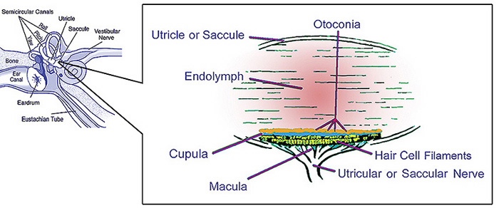
Caption = illustration of otolith organs showing detail ofutricle ,ococonia ,endolymph ,cupula ,macula ,hair cell filaments, andsaccular nerve
Caption2 =
MapPos = Saccule
System =
MeshName = Saccule+and+Utricle
MeshNumber = A09.246.631.909.625 |Introduction
The saccule is a bed of sensory cells situated in the inner ear. The saccule translates head movements into neural impulses which the brain can interpret. The saccule is sensitive to linear translations of the head, specifically movements up and down (think about moving on an elevator). When the head moves vertically, the sensory cells of the saccule are disturbed and the neurons connected to them begin transmitting impulses to the brain. These impulses travel along the vestibular portion of the eighth cranial nerve to the vestibular nuclei in the brainstem.
The vestibular system is important in maintaining
balance , orequilibrium . The vestibular system includes the saccule,utricle , and the threesemicircular canal s. Thevestibule is the name of the fluid-filled, membranous duct than contains these organs of balance. The vestibule is encased in thetemporal bone of the skull.Anatomy
The saccule, or sacculus, is the smaller of the two vestibular sacs. It is globular in form and lies in the
recessus sphæricus near the opening of thescala vestibuli of thecochlea . Its cavity does not directly communicate with that of theutricle . The anterior part of the saccule exhibits an oval thickening, the macula acustica sacculi, ormacula , to which are distributed the saccular filaments of the vestibular branch of thevestibulocochlear nerve , also known as theacoustic nerve orcranial nerve VIII .Within the macula are
hair cell s, each having ahair bundle on the apical aspect. The hair bundle is composed of a singlekinocilium and many (at least 70)stereocilia . Stereocilia are connected to mechanically-gated ion channels in the hair cell plasma membrane viatip link s. Supporting cells are interdigitate between hair cells and secrete theotolithic membrane , a thick, gelatinous layer of glycoprotein. Covering the surface of the otolithic membrane areotoliths , which are crystals of calcium carbonate. For this reason, the saccule is sometimes called an "otolithic organ."From the posterior wall of the saccule is given off a canal, the ductus endolymphaticus. This duct is joined by the ductus utriculosaccularis, and then passes along the aquæductus vestibuli and ends in a blind pouch (saccus endolymphaticus) on the posterior surface of the
petrous portion of thetemporal bone , where it is in contact with thedura mater .From the lower part of the saccule a short tube, the canalis reuniens of Hensen, passes downward and opens into the
ductus cochlearis near its vestibular extremity.Physiology
The saccule gathers sensory information to orient the body in space. It primarily gathers information about linear movement in the vertical plane. The structures that enable to saccule to gather this vestibular information are the
hair cell s. When the head tilts, the otolithic membrane slides over the hair cells in the direction of the tilt. This sliding bends the hair bundles of the hair cells, which causes stretching of the tip links and opening of the mechanically gated ion channels. Cations such as K+ rush into the hair cell cytosol, depolarizing it. Depolarization induces opening of voltage-gated Ca2+ channels at the basal aspect of the hair cell, which triggersexocytosis ofneurotransmitter to the vestibular neurons. These impulses are communicated via the vestibular nerve to vestibular nuclei in thebrainstem andmedulla . Impulses are also carried to thecerebellum via the inferiorcerebral peduncle s.ee also
*
Otolith
=AdditionalReferences
* Tortora, G. J. & Derrickson, B. (2006). "Principles of Anatomy and Physiology," 11th Edition. John Wiley and Sons, Inc.
External links
* [http://alpha.ipfw.edu/histo-embryo/wwwimagefolder/utricsac.jpgDiagram]

Wikimedia Foundation. 2010.