- Glossopharyngeal nerve
-
Nerve: Glossopharyngeal nerve 
Plan of upper portions of glossopharyngeal, vagus, and accessory nerves. 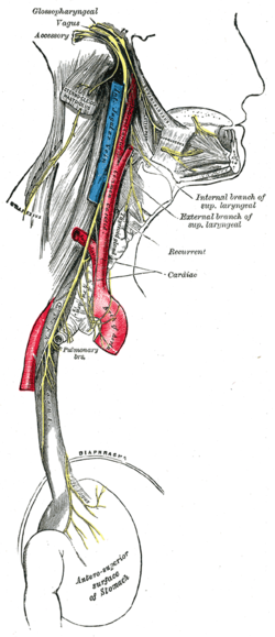
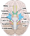
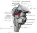
Course and distribution of the glossopharyngeal, vagus, and accessory nerves. (Label for glossopharyngeal is at upper right.) Latin nervus glossopharyngeus Gray's subject #204 906 Innervates stylopharyngeus To tympanic nerve MeSH Glossopharyngeal+Nerve Cranial Nerves CN I – Olfactory CN II – Optic CN III – Oculomotor CN IV – Trochlear CN V – Trigeminal CN VI – Abducens CN VII – Facial CN VIII – Vestibulocochlear CN IX – Glossopharyngeal CN X – Vagus CN XI – Spinal Accessory CN XII – Hypoglossal The glossopharyngeal nerve is the ninth (IX) of twelve pairs of cranial nerves (24 nerves total). It exits the brainstem out from the sides of the upper medulla, just rostral (closer to the nose) to the vagus nerve. The motor division of the glossopharyngeal nerve is derived from the basal plate of the embryonic medulla oblongata, while the sensory division originates from the cranial neural crest.
Contents
Functions
There are a number of functions of the glossopharyngeal nerve:
- It receives general sensory fibers (ventral trigeminothalamic tract) from the tonsils, the pharynx, the middle ear and the posterior 1/3 of the tongue.
- It receives special sensory fibers (taste) from the posterior one-third of the tongue.
- It receives visceral sensory fibers from the carotid bodies.
- It supplies parasympathetic fibers to the parotid gland via the otic ganglion.
- It supplies motor fibers to stylopharyngeus muscle, the only motor component of this cranial nerve.
- It contributes to the pharyngeal plexus.
Glossopharyngeal Overview
The glossopharyngeal nerve consists of five components with distinct functions: Branchial motor (special visceral efferent) - supplies the stylopharyngeus muscle. Visceral motor (general visceral efferent) provides parasympathetic innervation of the parotid gland. Visceral sensory (general visceral afferent) carries visceral sensory information from the carotid sinus and body. General sensory (general somatic afferent) provides general sensory information from the skin of the external ear, internal surface of the tympanic membrane, upper pharynx, and the posterior one-third of the tongue. Special sensory (special afferent) provides taste sensation from the posterior one-third of the tongue.
Overview of Branchial Motor Component
The branchial motor component of CN IX provides voluntary control of the stylopharyngeus muscle, which elevates the pharynx during swallowing and speech. Origin and Central Course - Branchial Motor Component. The branchial motor component originates from the nucleus ambiguus in the reticular formation of the medulla Rostral medulla. Fibers leaving the nucleus ambiguus travel anteriorly and laterally to exit the medulla, along with the other components of CN IX, between the olive and the inferior cerebellar peduncle. Intracranial Course - Branchial Motor Component. Upon emerging from the lateral aspect of the medulla the branchial motor component joins the other components of CN IX to exit the skull via the jugular foramen. The glossopharyngeal fibers travel just anterior to the cranial nerves X and XI, which also exit the skull via the jugular foramen. Extra-cranial course and final innervation. Upon exiting the skull the branchial motor fibers descend deep to the styloid process and wrap around the posterior border of the stylopharyngeus muscle before innervating it. Branchial motor component - voluntary control of the stylopharyngeus muscle. Signals for the voluntary movement of stylopharyngeus muscle originate in the pre-motor and motor cortex (in association with other cortical areas) and pass via the corticobulbar tract in the posterior limb of the internal capsule to synapse bilaterally on the ambiguus nuclei in the medulla.
Overview of visceral motor component
Parasympathetic component of the glossopharyngeal nerve that innervates the ipsilateral parotid gland.
Origin and central course
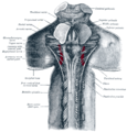
The preganglionic nerve fibers originate in the inferior salivatory nucleus of the rostral medulla and travel anteriorly and laterally to exit the brainstem between the olive and the inferior cerebellar peduncle with the other components of CN IX. Note: These neurons do not form a distinct nucleus visible on cross-section of the brainstem. The position indicated on the diagram is representative of the location of the cell bodies of these fibers.
Intracranial course
Upon emerging from the lateral aspect of the medulla, the visceral motor fibers join the other components of CN IX to enter the jugular foramen. Within the jugular foramen, there are two glossopharyngeal ganglia that contain nerve cell bodies that mediate general, visceral, and special sensation. The visceral motor fibers pass through both ganglia without synapsing and exit the inferior ganglion with CN IX general sensory fibers as the tympanic nerve. Before exiting the jugular foramen, the tympanic nerve enters the petrous portion of the temporal and ascends via the inferior tympanic canaliculus to the tympanic cavity. Within the tympanic cavity the tympanic nerve forms a plexus on the surface of the promontory of the middle ear to provide general sensation. The visceral motor fibers pass through this plexus and merge to become the lesser petrosal nerve. The lesser petrosal nerve re-enters and travels through the temporal bone to emerge in the middle cranial fossa just lateral to the greater petrosal nerve. It then proceeds anteriorly to exit the skull via the foramen ovale along with the mandibular component of CN V (V3).
Extra-cranial course and final innervations
Upon exiting the skull, the lesser petrosal nerve synapses in the otic ganglion, which is suspended from the mandibular nerve immediately below the foramen ovale. Postganglionic fibers from the otic ganglion travel with the auriculotemporal branch of CN V3 to enter the substance of the parotid gland.
Hypothalamic Influence
Fibers from the hypothalamus and olfactory system project via the dorsal longitudinal fasciculus to influence the output of the inferior salivatory nucleus. Examples include: 1) dry mouth in response to fear (mediated by the hypothalamus); 2) salivation in response to smelling food (mediated by the olfactory system)
Overview of visceral sensory component
This component of CN IX innervates the baroreceptors of the carotid sinus and chemoreceptors of the carotid body. Peripheral and intracranial course. Sensory fibers arise from the carotid sinus and carotid body at the bifurcation of the common carotid artery, ascend in the sinus nerve, and join the other components of CN IX at the inferior hypoglossal ganglion. The cell bodies of these neurons reside in the inferior ganglion. The central processes of these neurons enter the skull via the jugular foramen.
- Central course - visceral sensory component
- Once inside the skull, the visceral sensory fibers enter the lateral medulla between the olive and the inferior cerebellar peduncle and descend in the tractus solitarius to synapse in the caudal nucleus solitarius. From the nucleus solitarius, connections are made with several areas in the reticular formation and hypothalamus to mediate cardiovascular and respiratory reflex responses to changes in blood pressure, and serum concentrations of CO2 and O2.
Overview of general sensory component
This component of CN IX carries general sensory information (pain, temperature, and touch) from the skin of the external ear, internal surface of the tympanic membrane, the walls of the upper pharynx, and the posterior one-third of the tongue.
- Peripheral course
- Sensory fibers from the skin of the external ear initially travel with the auricular branch of CN X, while those from the middle ear travel in the tympanic nerve as discussed above (CN IX visceral motor section). General sensory information from the upper pharynx and posterior one-third of the tongue travel via the pharyngeal branches of CN IX. These peripheral processes have cell their cell body in either the superior or inferior glossopharyngeal ganglion.
Central course - general sensory component. The central processes of the general sensory neurons exit the glossopharyngeal ganglia and pass through the jugular foramen to enter the brainstem at the level of the medulla. Upon entering the medulla these fibers descend in the spinal trigeminal tract and synapse in the caudal spinal nucleus of the trigeminal.
- Central course - general sensory component
- Ascending secondary neurons originating from the spinal nucleus of CN V project to the contralateral ventral posteromedial (VPM) nucleus of the thalamus via the anterolateral system (ventral trigeminothalamic tract). Tertiary neurons from the thalamus project via the posterior limb of the internal capsule to the sensory cortex of the post-central gyrus.
Clinical correlation. The general sensory fibers of CN IX mediate the afferent limb of the pharyngeal reflex in which touching the back of the pharynx stimulates the patient to gag (i.e., the gag reflex). The efferent signal to the musculature of the pharynx is carried by the branchial motor fibers of the vagus nerve.
Overview of Special Sensory Component
The special sensory component of CN IX provides taste sensation from the posterior one-third of the tongue.
- Peripheral course
- Special sensory fibers from the posterior one-third of the tongue travel via the pharyngeal branches of CN IX to the inferior glossopharyngeal ganglion where their cell bodies reside.
- Central course - special sensory component
- The central processes of these neurons exit the inferior ganglion and pass through the jugular foramen to enter the brainstem at the level of the rostral medulla between the olive and inferior cerebellar peduncle. Upon entering the medulla, these fibers ascend in the tractus solitarius and synapse in the caudal nucleus solitarius. Taste fibers from CN VII and X also ascend and synapse here. Ascending secondary neurons originating in nucleus solitarius project bilaterally to the ventral posteromedial (VPM) nuclei of the thalamus via the central tegmental tract. Tertiary neurons from the thalamus project via the posterior limb of the internal capsule to the inferior one-third of the primary sensory cortex (the gustatory cortex of the parietal lobe).
Brainstem connections
The glossopharyngeal nerve is mostly sensory. The glossopharyngeal nerve also aids in tasting, swallowing and salivary secretions. Its superior and inferior (petrous) ganglia contain the cell bodies of pain fibers. It also projects into many different structures in the brainstem:
- Solitary nucleus: Taste from the posterior one-third of the tongue and information from carotid baroreceptors and carotid body chemoreceptors
- Spinal nucleus of the trigeminal nerve: Somatic sensory fibers from the middle ear
- Lateral Nucleus of Ala Cinerea: Visceral pain
- Nucleus ambiguus: The lower motor neurons for the stylopharyngeus muscle
- Inferior salivatory nucleus: Parasympathetic input to the parotid and mucous glands.
Path
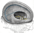
From the anterior portion of the medulla oblongata, the glossopharyngeal nerve passes laterally across or below the flocculus, and leaves the skull through the central part of the jugular foramen. From the superior and inferior ganglia in jugular foramen it has its own sheath of dura mater. The inferior ganglion on the inferior surface of petrous part of temporal is related with a triangular deppression into which the aqueduct of cochlea opens. On the inferior side, the glossopharyngeal nerve is lateral and anterior to the vagus nerve and accessory nerve.
In its passage through the jugular foramen (with X and XI), it passes between the internal jugular vein and internal carotid artery. It descends in front of the latter vessel, and beneath the styloid process and the muscles connected with it, to the lower border of the stylopharyngeus. It then curves forward, forming an arch on the side of the neck and lying upon the stylopharyngeus and middle pharyngeal constrictor muscle. From there, it passes under cover of the hyoglossus muscle, and is finally distributed to the palatine tonsil, the mucous membrane of the fauces and base of the tongue, and the mucous glands of the mouth
Branches
- Tympanic
- Stylopharyngeal
- Tonsillar
- Nerve to carotid sinus
- Branches to the posterior third of tongue
- Lingual branches
- A communicating branch to the Vagus nerve
Note: The glossopharyneal nerve contributes in the formation of the pharyngeal plexus along with the vagus nerve.
Testing the glossopharyngeal nerve
The integrity of the glossopharyngeal nerve may be evaluated by testing the patient's general sensation and that of taste on the posterior third of the tongue. The gag reflex can also be used to evaluate the glossphyaryngeal nerve, but also tests the vagus nerve, as only the afferent fibres involved in the reflex are carried by the glossopharyngeal nerve.
Additional images
External links
- NeuroNames hier-698
- MedEd at Loyola GrossAnatomy/h_n/cn/cn1/cn9.htm
- MedlinePlus Image 9350
- cranialnerves at The Anatomy Lesson by Wesley Norman (Georgetown University) (IX)
Nerves of head and neck: the cranial nerves and nuclei (TA A14.2.01, GA 9.855) olfactory (AON->I) optic (LGN->II) oculomotor
(ON, EWN->III)trochlear (TN->IV) no significant branchestrigeminal
(PSN, TSN, MN, TMN->V)abducens (AN->VI) no significant branchesfacial (FMN, SN, SSN->VII) near origininside
facial canalvestibulocochlear
(VN, CN->VIII)glossopharyngeal
(NA, ISN, SN->IX)before jugular fossaafter jugular fossavagus
(NA, DNVN, SN->X)before jugular fossaafter jugular fossaaccessory (NA, SAN->XI) hypoglossal (HN->XII) Categories:
Wikimedia Foundation. 2010.

