- Internal jugular vein
Infobox Vein
Name = Internal jugular vein
Latin = vena jugularis interna
GraySubject = 168
GrayPage = 648
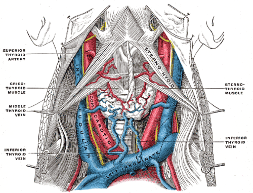
Caption = The fascia and middle thyroid veins. (Internal jugular visible at center left.)
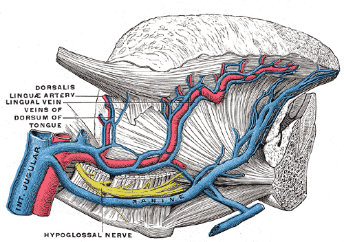
Caption2 = Veins of thetongue . Thehypoglossal nerve has been displaced downward in this preparation. (Internal jugular visible at bottom left.)
DrainsFrom =
Source = anterior facial
DrainsTo = brachiocephalic
Artery = internal carotid, common carotid
MeshName = Jugular+Veins
MeshNumber = A07.231.908.498
DorlandsPre = v_05
DorlandsSuf = 12850757
The internal jugular vein collects the blood from thebrain , the superficial parts of theface , and theneck .Path
It is directly continuous with the
sigmoid sinus , and begins in the posterior compartment of thejugular foramen , at the base of theskull .At its origin, it is somewhat dilated, and this dilatation is called the "superior bulb".
It also has a common trunk into which drains the anterior branch of the retro mandibular vein, the facial vein, and the lingual vein.
It runs down the side of the neck in a vertical direction, being at one end lateral to the
internal carotid artery , and then lateral to thecommon carotid , and at the root of the neck, it unites with thesubclavian vein to form thebrachiocephalic vein (innominate vein); a little above its termination is a second dilatation, the "inferior bulb".Above, it lies upon the
rectus capitis lateralis , behind the internal carotid artery and the nerves passing through the jugular foramen; lower down, the vein and artery lie upon the same plane, theglossopharyngeal andhypoglossal nerves passing forward between them; thevagus descends between and behind the vein and the artery in the same sheath (thecarotid sheath ), and the accessory runs obliquely backward, superficial or deep to the vein.At the root of the neck, the "right internal jugular vein" is a little distance from the
common carotid artery , and crosses the first part of thesubclavian artery , while the "left internal jugular vein" usually overlaps the common carotid artery.The left vein is generally smaller than the right, and each contains a pair of valves, which are placed about 2.5 cm above the termination of the vessel.
Clinical relevance
The jugular veins are relatively superficial and not protected by tissues such as
bone orcartilage . This makes them susceptible to damage. Due to the large volumes of blood that flow though the jugular veins, damage to the jugulars can quickly cause significant blood loss which can lead to hypovolæmic shock and then death if not treated.JVP
As there are no valves between the
right atrium of the heart and the internal jugular, blood can flow back into the internal jugular when the pressure in the atrium is sufficiently high. This can be seen from the outside, and allows one to estimate the pressure in the atrium. The pulsation seen is called thejugular venous pressure , or JVP. This is normally viewed with the patient at 45 degrees turning their head slightly away from the observer. The JVP can be raised in a number of conditions: [ [http://www.clinicalexam.com/pda/c_ref_jvp.htm Cardiovascular | Reference | JVP ] ]
*right ventricular failure (heart failure ),
*tricuspid stenosis
*tricuspid regurgitation
*cardiac tamponade The JVP can also be artificially raised by applying pressure to the liver (the
hepatojugular reflux ). This method is used to locate the JVP and distinguish it from the carotid pulse. Unlike the carotid pulse, the JVP is impalpable.Catheterization
As the internal jugular is large, central and relatively superficial, it is often used to place venous lines. Such a line may be inserted for several reasons, such as to accurately measure the central venous pressure or to administer fluids when a line in a peripheral vein would be unsuitable (such as during resuscitation when peripheral veins are hard to locate).
Because the internal jugular rarely varies in its location, it is easier to find than other veins. However, sometimes when a line is inserted the jugular is missed and other structures such as the
carotid artery or thevagus nerve (CN X) are punctured, causing damage to those structures.
=Additionalee also
*
jugular vein
*central venous catheter References
Wikimedia Foundation. 2010.