- Dura mater
-
Dura mater 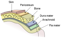
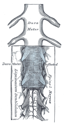
Meninges of the CNS Gray's subject #193 872 MeSH Dura+Mater The dura mater /ˈdjʊərə ˈmeɪtər/, or dura, is the outermost of the three layers of the meninges surrounding the brain and spinal cord. It is derived from Mesoderm. The other two meningeal layers are the pia mater and the arachnoid mater. The dura surrounds the brain and the spinal cord and is responsible for keeping in the cerebrospinal fluid. The name "dura mater" is derived from the Latin "hard mother" or "tough mother",[1] (translation of Arabic umm al-dimagh as-safiqa[2]) and is also referred to by the term "pachymeninx" (plural "pachymeninges").[3] The dura has been described as "tough and inflexible" and "leather-like".[3]
Contents
Layers and functions
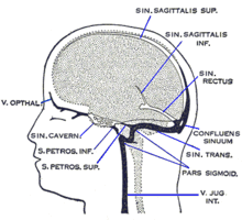
The dura mater has several functions and layers. The dura mater is a sac (aka thecal sac) that envelops the arachnoid mater. It surrounds and supports the dural sinuses (also called dural venous sinuses, cerebral sinuses, or cranial sinuses) and carries blood from the brain toward the heart.
The dura mater has two layers, or lamellae: The superficial layer, which serves as the skull's inner periosteum, called the endocranium; and a deep layer, the actual dura mater.
Dural folds and reflections
The dura separates into two layers at dural reflections (also known as dural folds), places where the inner dural layer is reflected as sheet-like protrusions into the cranial cavity. There are two main dural reflections:
- The tentorium cerebelli exists between and separates the cerebellum and brainstem from the occipital lobes of the cerebrum.[4]
- The falx cerebri, which separates the two hemispheres of the brain, is located in the longitudinal cerebral fissure between the hemispheres.[5]
Other two dural infoldings include the cerebellar falx and the sellar diaphragm
- The cerebellar falx (or Falx cerebelli) is a vertical dural infolding that lies inferior to the cerebellar tentorium in the posteiror part of the posterior cranial fossa. It partially separates the cerebellar hemispheres.
- The sellar diaphragm is the smallest dural infolding and is a circular sheet of dura that is suspended between the clinoid processes, forming a partial roof over the hypophysial fossa. The sellar diaphgram covers the pituitary gland in this fossa and has an aperture for passage of the infundibulum (pituitary stalk) and hypophysial veins.
Drainage
The two layers of dura mater run together throughout most of the skull. Where they separate, the gap between them is called a dural venous sinus. These sinuses drain blood and cerebrospinal fluid from the brain and empty into the internal jugular vein.
They drain via the arachnoid villi, which are outgrowths of the arachnoid mater (the middle meningeal layer) that extend into the venous sinuses. These villi act as one-way valves.
Meningeal veins, which course through the dura mater, and bridging veins, which drain the underlying neural tissue and puncture the dura mater, empty into these dural sinuses.
Clinical significance
Many medical conditions involve the dura mater. A subdural hematoma occurs when there is an abnormal collection of blood between the dura and the arachnoid, usually as a result of torn bridging veins secondary to head trauma. An epidural hematoma is a collection of blood between the dura and the inner surface of the skull, and is usually due to arterial bleeding.
In 2011, Scali et al., discovered a connection of soft tissue from the rectus capitis posterior major to the cervical dura mater. Various clinical manifestations may be linked to this anatomical relationship such as headaches, trigeminal neuralgia and other symptoms that involved the cervical dura (Spine 2011).[6] The rectus capitis posterior minor has a similar attachment as described by Hack et al. in 1995.
The dura-muscular, dura-ligamentous connections in the upper cervical spine and occipital areas may provide anatomic and physiologic answers to the cause of the cervicogenic headache. This proposal would further explain manipulation's efficacy in the treatment of cervicogenic headache.[7]
Blood supply
The middle meningeal artery supplies most of the blood for the dura mater, though the meningeal branches of the posterior and anterior ethmoidal artery also contribute.
Innervation
Sensory innervation of the supratentorial dura mater is via small meningeal branches of the trigeminal nerve (V1, V2 and V3).[8] The innervation for the infratentorial dura mater are upper cervical nerves.
The American Red Cross and some other agencies accepting blood donations consider dura mater transplants, along with receipt of pituitary-derived growth hormone, a risk factor due to concerns about Creutzfeldt-Jakob disease.[9]
Dural ectasia is the enlargement of the dura and is common in connective tissue disorders, such as Marfan Syndrome and Ehlers-Danlos Syndrome.
Spontaneous Cerebrospinal Fluid Leak is the fluid and pressure loss of spinal fluid due to holes in the dura mater.
References
- ^ http://www.medterms.com/script/main/art.asp?articlekey=32513
- ^ http://www.etymonline.com/index.php?term=dura+mater
- ^ a b http://www.medterms.com/script/main/art.asp?articlekey=32512
- ^ Shepherd S. 2004. "Head Trauma." Emedicine.com.
- ^ Vinas FC and Pilitsis J. 2004. "Penetrating Head Trauma." Emedicine.com.
- ^ Frank Scali, Eric S. Marsili, Matt E. Pontell. "Anatomical Connection Between the Rectus Capitis Posterior Major and the Dura Mater". Spine. http://journals.lww.com/spinejournal/Abstract/publishahead/Anatomical_Connection_Between_the_Rectus_Capitis.99006.aspx.
- ^ Gary D. Hack, Peter Ratiu, John P. Kerr, Gwendolyn F. Dunn, Mi Young Toh. "Visualization of the Muscle-Dural Bridge in the Visible Human Female Data Set". The Visible Human Project, National Library of Medicine. http://www.nlm.nih.gov/research/visible/vhp_conf/hack2/hack2.htm.
- ^ 'Gray's Anatomy for Students' 2005, Drake, Vogl and Mitchell, Elsevier
- ^ Red Cross: http://www.redcross.org/services/biomed/0,1082,0_553_,00.html
Additional images
External links
- Dura+mater at eMedicine Dictionary
Anatomy: meninges of the brain and medulla spinalis (TA A14.1.01, GA 9.749/9.872) Layers Dura materArachnoid granulation · Arachnoid trabeculae
Subarachnoid cisterns: Cisterna magna · Pontine cistern · Interpeduncular cistern · Chiasmatic · Lateral cerebral fossa · Of great cerebral vein · Of lamina terminalisTela chorioidea (Tela chorioidea of third ventricle, Tela chorioidea of fourth ventricle) · Choroid plexusCombinedSpaces Anatomy of torso (primarily): the spinal cord (TA 14.1.02, GA 9.749) External, dorsal Posterior median sulcus · Posterolateral sulcusGrey matter/
Rexed laminaeI–VI: Posterior hornI: Marginal nucleus · II: Substantia gelatinosa of Rolando · III+IV: Nucleus proprius · Spinal lamina V · Spinal lamina VIVII: Lateral hornVIII–IX: Anterior hornX: OtherWhite matter somatic/
ascending
(blue)Posterior/PCML: touch: Gracile · Cuneate
Lateral: proprioception: Spinocerebellar (Dorsal, Ventral) · pain/temp: Spinothalamic (Lateral, Anterior) · Posterolateral (Lissauer) · Spinotectal
Spinoreticular tract · Spino-olivary tractmotor/
descending
(red)Lateral: Corticospinal (Lateral) · Ep (Rubrospinal, Olivospinal)
Anterior: Corticospinal (Anterior) · Ep (Vestibulospinal, Reticulospinal, Tectospinal)bothExternal, ventral Anterior median fissure · Anterolateral sulcusExternal, general Categories:
Wikimedia Foundation. 2010.

