- Vertebral artery
Infobox Artery
Name = Vertebral artery
Latin = arteria vertebralis
GraySubject = 148
GrayPage = 578
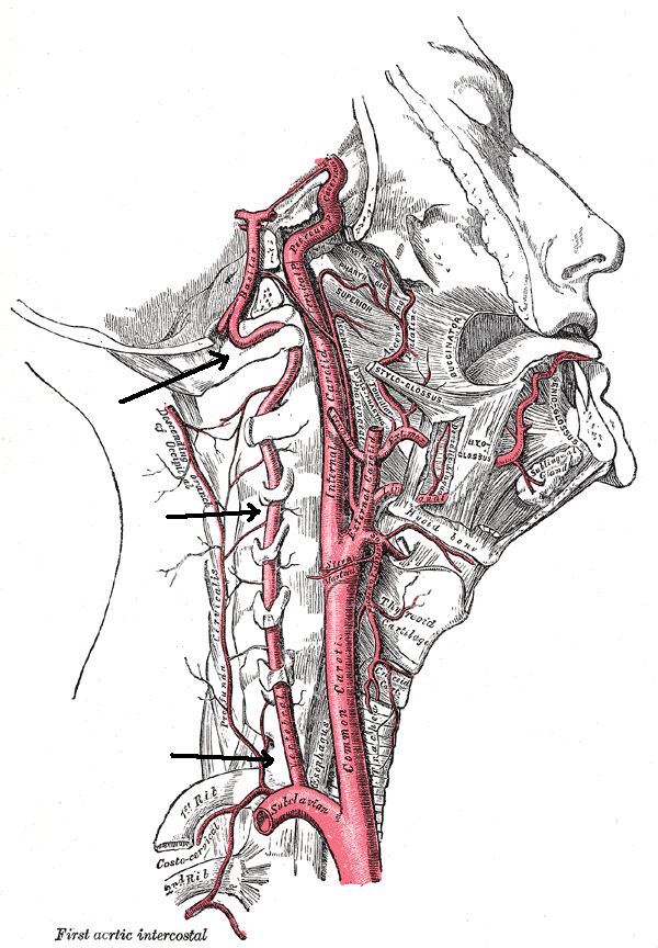
Caption = Arteries of the neck. The vertebral arteries arise from the subclavian arteries and join to form thebasilar artery . It is ponted out, centermost of the three vertical arteries.
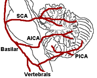
Caption2 = The three major arteries of the cerebellum: the SCA, AICA, and PICA. (Vertebrals labeled at bottom.)
BranchFrom = subclavian arteries
BranchTo = Meningeal branches
Posterior spinal
Anterior spinal
PICABasilar artery
Vein =vertebral vein
Supplies =
MeshName = Subclavian+Artery
MeshNumber = A07.231.114.839
DorlandsPre = a_61
DorlandsSuf = 12156505
The vertebral arteries are branches of the subclavian arteries.The two vertebral arteries and the
basilar artery are sometimes together called the vertebrobasilar system, which supplies blood to the posterior part ofcircle of Willis and anastomoses with blood supplied to the anterior part of the circle of Willis from the carotid arteries.Cervical
They arise, one on each side of the body, then enter deep to the transverse process of the level of the 6th
cervical vertebrae (C6).It then proceeds superiorly, in the
transverse foramen (foramen transversarium) of eachcervical vertebra until C1.This path is largely parallel to, but distinct from, the route of the
carotid artery ascending through the neck.At the C1 level the vertebral arteries travel across the posterior arch of the atlas before entering the
foramen magnum .Cranial
Inside the skull, the two vertebral arteries join up to form the
basilar artery at the base of themedulla oblongata .The
basilar artery is the main blood supply to thebrainstem and connects to theCircle of Willis to potentially supply the rest of the brain if there is compromise to one of the carotids.At each cervical level, the vertebral artery sends branches to the surrounding musculature via
anterior spinal arteries .Division into four parts
The vertebral artery may be divided into four parts:
First part
The first part runs upward and backward between the
Longus colli and theScalenus anterior .In front of it are the
internal jugular andvertebral vein s, and it is crossed by theinferior thyroid artery ; the left vertebral is crossed by thethoracic duct also.Behind it are the transverse process of the seventh
cervical vertebra , thesympathetic trunk and itsinferior cervical ganglion .econd part
The second part runs upward through the
foramina in thetransverse processes of the upper six cervical vertebræ, and is surrounded by branches from the inferior cervical sympathetic ganglion and by a plexus of veins which unite to form the vertebral vein at the lower part of the neck.It is situated in front of the trunks of the cervical nerves, and pursues an almost vertical course as far as the
transverse process of the atlas , above which it runs upward and lateralward to the foramen in the transverse process of the atlas.Third part
The third part issues from the latter foramen on the medial side of the
Rectus capitis lateralis , and curves backward behind thesuperior articular process of the atlas , the anterior ramus of the first cervical nerve being on its medial side; it then lies in the groove on the upper surface of the posterior arch of the atlas, and enters the vertebral canal by passing beneath theposterior atlantoöccipital membrane .This part of the artery is covered by the
Semispinalis capitis and is contained in thesuboccipital triangle —a triangular space bounded by theRectus capitis posterior major , theObliquus superior , and theObliquus inferior .The first cervical or
suboccipital nerve lies between the artery and theposterior arch of the atlas.Fourth part
The fourth part pierces the
dura mater and inclines medialward to the front of themedulla oblongata ; it is placed between thehypoglossal nerve and the anterior root of the first cervical nerve and beneath the first digitation of theligamentum denticulatum .At the lower border of the
pons it unites with the vessel of the opposite side to form thebasilar artery .Asymmetry
The left vertebral artery is usually larger and carries more blood. [cite journal |author= Albayrak, Ramazan|title=Doppler sonography evaluation of flow velocity and volume of the extracranial internal carotid and vertebral arteries in healthy adults |journal=J Clin Ultrasound |volume=35 |issue=1 |pages=27–33 |year=2007 |pmid=17149761|doi=10.1002/jcu.20301]
ee also
*
Arcuate foramen References
=AdditionalExternal links
*
*
*
*
* (NormanAnatomyFig|vertebralcolumnfromleftweb)
*
*
Wikimedia Foundation. 2010.