Dorsal column nuclei

Dorsal column nuclei
{{{Name}}}
Gray693.png
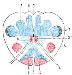
Present at the junction between the spinal cord and medulla oblongata, the dorsal column nuclei consist of paired gracile and cuneate nuclei (labels 6 and 7, respectively).
System Somatosensory system

In neuroanatomy, the dorsal column nuclei are a pair of nuclei in the brainstem. The name refers collectively to the cuneate nucleus and gracile nucleus, which are present at the junction between the spinal cord and the medulla oblongata. Both nuclei contain secondary neurons of the dorsal column-medial lemniscus pathway, which carries fine touch and proprioceptive information from the body to the brain.

The gracile nucleus is medial to the cuneate nucleus; its neurons receive afferent input from dorsal root ganglion sensory neurons subserving the lower trunk and limbs, while neurons of the cuneate nucleus receive connections from dorsal root neurons innervating the upper body. Neurons of the dorsal column nuclei send axons that form the internal arcuate fibers, decussating (crossing to the opposite side) to form the medial lemniscus, ultimately synapsing with third-order neurons of the thalamus.

Because each nucleus contains a large population of neurons, the dorsal column nuclei give rise to characteristic bumps or tubercles when viewing the posterior side of the intact brainstem. In particular, the cuneate nucleus gives rise to the cuneate tubercle, while the gracile nucleus gives rise to the gracile tubercle.


Wikimedia Foundation. 2010.

Игры ⚽ Поможем решить контрольную работу

Look at other dictionaries:

  • dorsal column nuclei — the nucleus cuneatus and nucleus gracilis, which are at the upper end of the dorsal column of the spinal cord. Called also posterior column nuclei …   Medical dictionary

  • posterior column nuclei — dorsal column nuclei …   Medical dictionary

  • Cochlear nuclei — Brain: Cochlear nuclei Dissection of brain stem. Dorsal view. ( Cochlear nucleus is labeled on left, fifth from the bottom.) …   Wikipedia

  • column — 1. An anatomic part or structure in the form of a pillar or cylindric funiculus. SEE ALSO: fascicle. 2. A vertical object (usually cylindrical), mass, or formation. SYN: columna [TA]. [L. columna] …   Medical dictionary

  • Column of Clarke — Infobox Anatomy Name = PAGENAME Latin = nucleus thoracicus posterior, nucleus dorsalis GraySubject = 185 GrayPage = 758 Caption = Diagram showing a few of the connections of afferent (sensory) fibers of the posterior root with the efferent fibers …   Wikipedia

  • Posterior column-medial lemniscus pathway — Originating in peripheral sensory receptors, the posterior column medial lemniscus pathway transmits fine touch and conscious proprioceptive information to the brain. Latin via column …   Wikipedia

  • Spinal cord — Latin medulla spinalis The spinal cord nested in the vertebral column …   Wikipedia

  • List of anatomical topics — This page aims to list articles related to anatomy. This list is not necessarily complete or up to date if you see an article that should be here but isn t (or one that shouldn t be here but is), please update the page accordingly. NOTOC A… …   Wikipedia

  • Medulla oblongata — Brain: Medulla oblongata labeled at bottom left …   Wikipedia

  • Trigeminal nerve — Nerve: Trigeminal nerve Trigeminal nerve, shown in yellow …   Wikipedia

Share the article and excerpts

Direct link
Do a right-click on the link above
and select “Copy Link”